Explore
 Validate
 Learn
Learn Western blot
 Immunocytochemistry
Antibody data
- Antibody Data
- Antigen structure
- References [0]
- Comments [0]
- Validations
- Western blot [1]
- Immunocytochemistry [1]
- Immunohistochemistry [1]
Submit
Validation data
Reference
Comment
Report error
- Product number
- AMAb91220 - Provider product page

- Provider
- Atlas Antibodies
- Proper citation
- Atlas Antibodies Cat#AMAb91220, RRID:AB_2665849
- Product name
- Anti-CDH2
- Antibody type
- Monoclonal
- Description
- Monoclonal Antibody against Human CDH2, Clone ID: CL3716, Gene description: cadherin 2, type 1, N-cadherin (neuronal), Alternative Gene Names: CD325, CDHN, NCAD, Validated applications: WB, IHC, ICC, Uniprot ID: P19022, Storage: Store at +4°C for short term storage. Long time storage is recommended at -20°C.
- Reactivity
- Human
- Host
- Mouse
- Conjugate
- Unconjugated
- Isotype
- IgG
- Antibody clone number
- CL3716
- Vial size
- 100 µl
- Concentration
- 1.0 mg/ml
- Storage
- Store at +4°C for short term storage. Long time storage is recommended at -20°C.
- Handling
- The antibody solution should be gently mixed before use.
No comments: Submit comment
Enhanced validation
- Submitted by
- Atlas Antibodies (provider)
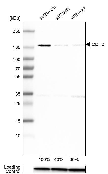
- Enhanced method
- Genetic validation
- Main image

- Experimental details
- Western blot analysis in U-251MG cells transfected with control siRNA, target specific siRNA probe #1 and #2, using Anti-CDH2 antibody. Remaining relative intensity is presented. Loading control: Anti-GAPDH.
- Sample type
- Human
- Protocol
- Protocol
Supportive validation
- Submitted by
- Atlas Antibodies (provider)
- Main image
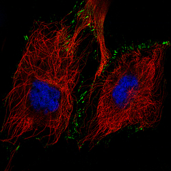
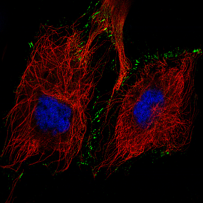
- Experimental details
- Immunofluorescence staining of U-251 cells using the Anti-CDH2 monoclonal antibody, showing specific staining in the plasma membrane and cell junctions in green. Microtubule- and nuclear probes are visualized in red and blue, respectively (where available).
- Sample type
- Human
Supportive validation
- Submitted by
- Atlas Antibodies (provider)
- Enhanced method
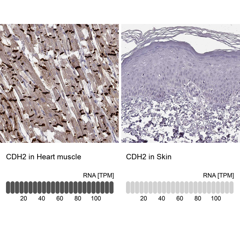
- Orthogonal validation
- Main image
- Experimental details
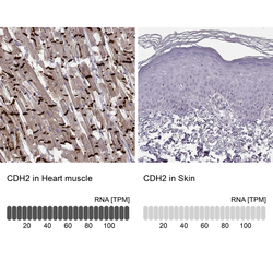
- Immunohistochemistry analysis in human heart muscle and skin tissues using AMAb91220 antibody. Corresponding CDH2 RNA-seq data are presented for the same tissues.
- Sample type
- Human
- Protocol
- Protocol