Explore
 Validate
 Learn
Learn Western blot
 Immunocytochemistry
 Immunoprecipitation
Antibody data
- Antibody Data
- Antigen structure
- References [5]
- Comments [0]
- Validations
- Immunocytochemistry [2]
- Immunohistochemistry [1]
- Other assay [1]
Submit
Validation data
Reference
Comment
Report error
- Product number
- PA5-19486 - Provider product page

- Provider
- Invitrogen Antibodies
- Product name
- N-cadherin Polyclonal Antibody
- Antibody type
- Polyclonal
- Antigen
- Synthetic peptide
- Description
- Heat mediated antigen retrieval recommended prior to tissue staining. This antibody is predicted to react with chicken and cow based on sequence homology.
- Reactivity
- Human, Mouse, Rat
- Host
- Rabbit
- Isotype
- IgG
- Vial size
- 100 μg
- Concentration
- 0.8 mg/mL
- Storage
- -20°C or -80°C if preferred
Submitted references Neonatal Hyperoxia Downregulates Claudin-4, Occludin, and ZO-1 Expression in Rat Kidney Accompanied by Impaired Proximal Tubular Development.
H19-Dependent Transcriptional Regulation of β3 and β4 Integrins Upon Estrogen and Hypoxia Favors Metastatic Potential in Prostate Cancer.
VCAM-1 induces signals that stimulate ZO-1 serine phosphorylation and reduces ZO-1 localization at lung endothelial cell junctions.
ZEB1 Regulates Multiple Oncogenic Components Involved in Uveal Melanoma Progression.
Cisplatin promotes mesenchymal-like characteristics in osteosarcoma through Snail.
Xu X, Zhang X, Gao L, Liu C, You K
Oxidative medicine and cellular longevity 2020;2020:2641461
Oxidative medicine and cellular longevity 2020;2020:2641461
H19-Dependent Transcriptional Regulation of β3 and β4 Integrins Upon Estrogen and Hypoxia Favors Metastatic Potential in Prostate Cancer.
Bacci L, Aiello A, Ripoli C, Loria R, Pugliese D, Pierconti F, Rotili D, Strigari L, Pinto F, Bassi PF, Mai A, Grassi C, Pontecorvi A, Falcioni R, Farsetti A, Nanni S
International journal of molecular sciences 2019 Aug 17;20(16)
International journal of molecular sciences 2019 Aug 17;20(16)
VCAM-1 induces signals that stimulate ZO-1 serine phosphorylation and reduces ZO-1 localization at lung endothelial cell junctions.
Abdala-Valencia H, Kountz TS, Marchese ME, Cook-Mills JM
Journal of leukocyte biology 2018 Jul;104(1):215-228
Journal of leukocyte biology 2018 Jul;104(1):215-228
ZEB1 Regulates Multiple Oncogenic Components Involved in Uveal Melanoma Progression.
Chen Y, Lu X, Montoya-Durango DE, Liu YH, Dean KC, Darling DS, Kaplan HJ, Dean DC, Gao L, Liu Y
Scientific reports 2017 Mar 3;7(1):45
Scientific reports 2017 Mar 3;7(1):45
Cisplatin promotes mesenchymal-like characteristics in osteosarcoma through Snail.
Fang S, Yu L, Mei H, Yang J, Gao T, Cheng A, Guo W, Xia K, Liu G
Oncology letters 2016 Dec;12(6):5007-5014
Oncology letters 2016 Dec;12(6):5007-5014
No comments: Submit comment
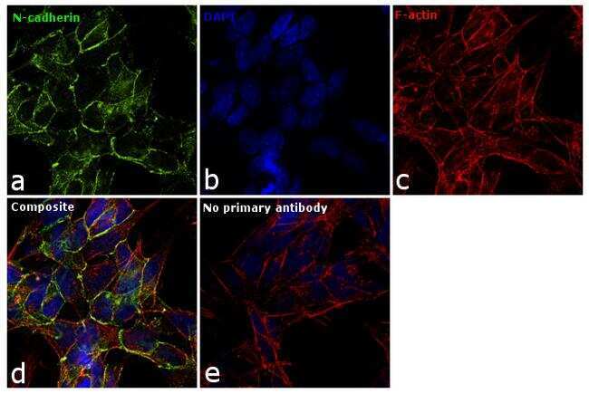
Supportive validation
- Submitted by
- Invitrogen Antibodies (provider)
- Main image
- Experimental details
- Immunofluorescence analysis of N-cadherin was performed using 90% confluent log phase SH-SY5Y cells. The cells were fixed with 4% paraformaldehyde for 10 minutes, permeabilized with 0.1% Triton™ X-100 for 15 minutes, and blocked with 1% BSA for 1 hour at room temperature. The cells were labeled with N-cadherin Monoclonal Antibody (Product # PA5-19486) at 5 µg/mL in 0.1% BSA, incubated at 4 degree Celsius overnight and then labeled with Goat anti-Rabbit IgG (H+L) Superclonal™ Secondary Antibody, Alexa Fluor® 488 conjugate (Product # A27034) at a dilution of 1:2000 for 45 minutes at room temperature (Panel a: green). Nuclei (Panel b: blue) were stained with SlowFade® Gold Antifade Mountant with DAPI (Product # S36938). F-actin (Panel c: red) was stained with Rhodamine Phalloidin (Product # R415, 1:300). Panel d represents the merged image showing membranous localization. Panel e represents control cells with no primary antibody to assess background. The images were captured at 60X magnification.
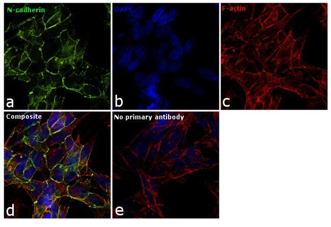
- Submitted by
- Invitrogen Antibodies (provider)
- Main image

- Experimental details
- Immunofluorescence analysis of N-cadherin was performed using 90% confluent log phase SH-SY5Y cells. The cells were fixed with 4% paraformaldehyde for 10 minutes, permeabilized with 0.1% Triton™ X-100 for 15 minutes, and blocked with 1% BSA for 1 hour at room temperature. The cells were labeled with N-cadherin Monoclonal Antibody (Product # PA5-19486) at 5 µg/mL in 0.1% BSA, incubated at 4 degree Celsius overnight and then labeled with Goat anti-Rabbit IgG (Heavy Chain) Superclonal™ Secondary Antibody, Alexa Fluor® 488 conjugate (Product # A27034) at a dilution of 1:2000 for 45 minutes at room temperature (Panel a: green). Nuclei (Panel b: blue) were stained with SlowFade® Gold Antifade Mountant with DAPI (Product # S36938). F-actin (Panel c: red) was stained with Rhodamine Phalloidin (Product # R415, 1:300). Panel d represents the merged image showing membranous localization. Panel e represents control cells with no primary antibody to assess background. The images were captured at 60X magnification.
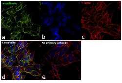
Supportive validation
- Submitted by
- Invitrogen Antibodies (provider)
- Main image
- Experimental details
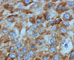
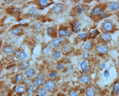
- Immunohistochemical (formalin-fixed, paraffin-embedded) staining of Human Liver Cancer tissue using Product # PA5-19486, anti-N Cadherin antibody. Primary antibody was used at a concentration of 1 µg/mL and exposed for 15 mins at room temp. The sample was pretreated using heat mediated antigen retrieval with Sodium Citrate Buffer (pH6/20mins). The detection method was a HRP conjugated polymer, DAB chromogen and the sample was counterstained with haematoxylin and mounted with DPX.
Supportive validation
- Submitted by
- Invitrogen Antibodies (provider)
- Main image
- Experimental details
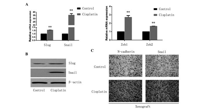
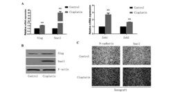
- Figure 2. Cisplatin promotes EMT-TFs in osteosarcoma. (A) The relative expression of EMT-TFs, including Snail/Slug and Zeb1/2 were observed to be significantly upregulated in the cisplatin treated cells by quantitative polymerase chain reaction. **P