Explore
 Validate
 Learn
Learn Western blot
Antibody data
- Antibody Data
- Antigen structure
- References [5]
- Comments [0]
- Validations
- Western blot [1]
- ELISA [1]
- Immunocytochemistry [1]
- Immunohistochemistry [1]
- Flow cytometry [1]
Submit
Validation data
Reference
Comment
Report error
- Product number
- AF6426 - Provider product page

- Provider
- R&D Systems
- Product name
- Human/Mouse/Rat N-Cadherin Antibody
- Antibody type
- Polyclonal
- Description
- Antigen Affinity-purified. Detects human, mouse, and rat N-Cadherin in Western blots. In direct ELISA, less than 10% cross-reactivity with recombinant human (rh) E-Cadherin, and rhR-Cadherin is observed.
- Reactivity
- Human, Mouse, Rat
- Host
- Sheep
- Conjugate
- Unconjugated
- Antigen sequence
P19022- Isotype
- IgG
- Vial size
- 100 ug
- Concentration
- LYOPH
- Storage
- Use a manual defrost freezer and avoid repeated freeze-thaw cycles. 12 months from date of receipt, -20 to -70 °C as supplied. 1 month, 2 to 8 °C under sterile conditions after reconstitution. 6 months, -20 to -70 °C under sterile conditions after reconstitution.
Submitted references Organoid single cell profiling identifies a transcriptional signature of glomerular disease.
MicroRNA-149-5p regulates blood-brain barrier permeability after transient middle cerebral artery occlusion in rats by targeting S1PR2 of pericytes.
BDNF/TrkB axis activation promotes epithelial-mesenchymal transition in idiopathic pulmonary fibrosis.
MMP20 modulates cadherin expression in ameloblasts as enamel develops.
In vivo biomarker expression patterns are preserved in 3D cultures of Prostate Cancer.
Harder JL, Menon R, Otto EA, Zhou J, Eddy S, Wys NL, O'Connor C, Luo J, Nair V, Cebrian C, Spence JR, Bitzer M, Troyanskaya OG, Hodgin JB, Wiggins RC, Freedman BS, Kretzler M, European Renal cDNA Bank (ERCB), Nephrotic Syndrome Study Network (NEPTUNE)
JCI insight 2019 Jan 10;4(1)
JCI insight 2019 Jan 10;4(1)
MicroRNA-149-5p regulates blood-brain barrier permeability after transient middle cerebral artery occlusion in rats by targeting S1PR2 of pericytes.
Wan Y, Jin HJ, Zhu YY, Fang Z, Mao L, He Q, Xia YP, Li M, Li Y, Chen X, Hu B
FASEB journal : official publication of the Federation of American Societies for Experimental Biology 2018 Jun;32(6):3133-3148
FASEB journal : official publication of the Federation of American Societies for Experimental Biology 2018 Jun;32(6):3133-3148
BDNF/TrkB axis activation promotes epithelial-mesenchymal transition in idiopathic pulmonary fibrosis.
Cherubini E, Mariotta S, Scozzi D, Mancini R, Osman G, D'Ascanio M, Bruno P, Cardillo G, Ricci A
Journal of translational medicine 2017 Sep 22;15(1):196
Journal of translational medicine 2017 Sep 22;15(1):196
MMP20 modulates cadherin expression in ameloblasts as enamel develops.
Guan X, Bartlett JD
Journal of dental research 2013 Dec;92(12):1123-8
Journal of dental research 2013 Dec;92(12):1123-8
In vivo biomarker expression patterns are preserved in 3D cultures of Prostate Cancer.
Windus LC, Kiss DL, Glover T, Avery VM
Experimental cell research 2012 Nov 15;318(19):2507-19
Experimental cell research 2012 Nov 15;318(19):2507-19
No comments: Submit comment
Supportive validation
- Submitted by
- R&D Systems (provider)
- Main image
- Experimental details
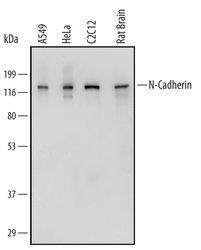
- Detection of Human, Mouse, and Rat N-Cadherin by Western Blot. Western blot shows lysates of A549 human lung carcinoma cell line, HeLa human cervical epithelial carcinoma cell line, C2C12 mouse myoblast cell line, and rat brain tissue. PVDF Membrane was probed with 0.5 µg/mL of Sheep Anti-Human/Mouse/Rat N-Cadherin Antigen Affinity-purified Polyclonal Antibody (Catalog # AF6426) followed by HRP-conjugated Anti-Sheep IgG Secondary Antibody (Catalog # HAF016). A specific band was detected for N-Cadherin at approximately 130 kDa (as indicated). This experiment was conducted under reducing conditions and using Immunoblot Buffer Group 1.

Supportive validation
- Submitted by
- R&D Systems (provider)
- Main image
- Experimental details
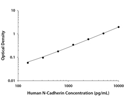
- Human N-Cadherin ELISA Standard Curve. Recombinant Human N-Cadherin protein was serially diluted 2-fold and captured by Mouse Anti-Human N-Cadherin Monoclonal Antibody (Catalog # MAB13883) coated on a Clear Polystyrene Microplate (Catalog # DY990). Sheep Anti-Human/Mouse/Rat N-Cadherin Antigen Affinity-purified Polyclonal Antibody (Catalog # AF6426) was biotinylated and incubated with the protein captured on the plate. Detection of the standard curve was achieved by incubating Streptavidin-HRP (Catalog # DY998) followed by Substrate Solution (Catalog # DY999) and stopping the enzymatic reaction with Stop Solution (Catalog # DY994).

Supportive validation
- Submitted by
- R&D Systems (provider)
- Main image
- Experimental details
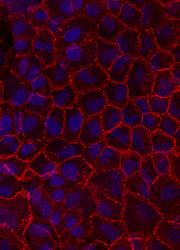
- N-Cadherin in A549 Human Cell Line. N-Cadherin was detected in immersion fixed A549 human lung carcinoma cell line using Sheep Anti-Human/Mouse/Rat N-Cadherin Affinity-purified Polyclonal Antibody (Catalog # AF6426) at 10 µg/mL for 3 hours at room temperature. Cells were stained using the NorthernLights™ 557-conjugated Anti-Sheep IgG Secondary Antibody (red; Catalog # NL010) and counterstained with DAPI (blue). Specific staining was localized to cell surfaces. View our protocol for Fluorescent ICC Staining of Cells on Coverslips.

Supportive validation
- Submitted by
- R&D Systems (provider)
- Main image
- Experimental details
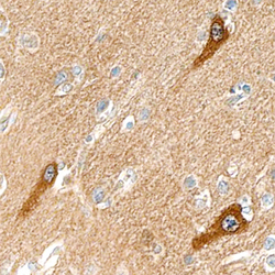
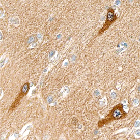
- N-Cadherin in Human Brain. N-Cadherin was detected in immersion fixed paraffin-embedded sections of human brain (cortex) using Sheep Anti-Human/Mouse/Rat N-Cadherin Antigen Affinity-purified Polyclonal Antibody (Catalog # AF6426) at 15 µg/mL overnight at 4 °C. Tissue was stained using the Anti-Sheep HRP-DAB Cell & Tissue Staining Kit (brown; Catalog # CTS019) and counterstained with hematoxylin (blue). Specific staining was localized to neuronal cell bodies and processes. View our protocol for Chromogenic IHC Staining of Paraffin-embedded Tissue Sections.
Supportive validation
- Submitted by
- R&D Systems (provider)
- Main image
- Experimental details
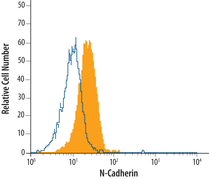
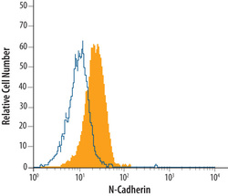
- Detection of N-Cadherin in HeLa Human Cell Line by Flow Cytometry. HeLa human cervical epithelial carcinoma cell line was stained with Sheep Anti-Human/Mouse/Rat N-Cadherin Antigen Affinity-purified Polyclonal Antibody (Catalog # AF6426, filled histogram) or Sheep IgG control antibody (Catalog # 5-001-A, open histogram), followed by NorthernLights™ 557-conjugated Anti-Sheep IgG Secondary Antibody (Catalog # NL010).