Explore
 Validate
 Learn
Learn Western blot
 Immunocytochemistry
Antibody data
- Antibody Data
- Antigen structure
- References [0]
- Comments [0]
- Validations
- Western blot [1]
- Immunohistochemistry [1]
- Flow cytometry [5]
Submit
Validation data
Reference
Comment
Report error
- Product number
- NB200-585-0.1mg - Provider product page

- Provider
- Novus Biologicals
- Proper citation
- Novus Cat#NB200-585-0.1mg, RRID:AB_10000865
- Product name
- Mouse Monoclonal TfR (Transferrin R) Antibody
- Antibody type
- Monoclonal
- Description
- Protein G purified. Labels dividing cells of various types but also binds to a number of non-dividing normal tissues and endothelium of brain capillaries.
- Reactivity
- Mouse, Rat
- Host
- Mouse
- Isotype
- IgG
- Vial size
- 0.1 mg
- Concentration
- 1.0 mg/ml
- Storage
- Store at 4C short term. Aliquot and store at -20C long term. Avoid freeze-thaw cycles.
No comments: Submit comment
Supportive validation
- Submitted by
- Novus Biologicals (provider)
- Main image

- Experimental details
- Western Blot: TfR (Transferrin R) Antibody (OX26) [NB200-585] - Total protein from rat adrenal gland cells (PC12) and rat placenta was separated on a 7.5% gel by SDS-PAGE, transferred to PVDF membrane and blocked in 5% non-fat milk in TBST. The membrane was probed with 2 ug/ml anti-CD71 in 1% milk, and detected with an anti-mouse HRP secondary antibody using chemiluminescence.

Supportive validation
- Submitted by
- Novus Biologicals (provider)
- Main image

- Experimental details
- Immunohistochemistry-Paraffin: TfR (Transferrin R) Antibody (OX26) [NB200-585] - Analysis of FFPE mouse spleen using TfR (OX26) antibody at 1:50 on a Bond Rx autostainer (Leica Biosystems). The assay involved 20 minutes of heat induced antigen retrieval (HIER) using 10mM sodium citrate buffer (pH 6.0) and endogenous peroxidase quenching with peroxide block. The sections were incubated with primary antibody for 30 minutes and Bond Polymer Refine Detection (Leica Biosystems) with DAB was used for signal development followed by counterstaining with hematoxylin. Whole slide scanning and capturing of representative images was performed using Aperio AT2 (Leica Biosystems). Plasma membrane staining was observed. Staining was performed by Histowiz.

Supportive validation
- Submitted by
- Novus Biologicals (provider)
- Main image

- Experimental details
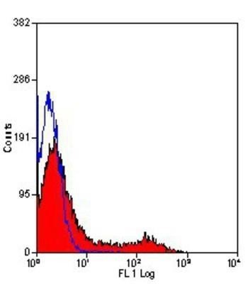
- Flow Cytometry: TfR (Transferrin R) Antibody (OX26) [NB200-585] - Staining of rat spleen lymphocytes with mouse anti rat CD71.

- Submitted by
- Novus Biologicals (provider)
- Main image

- Experimental details
- Flow (Intracellular): TfR (Transferrin R) Antibody (OX26) [NB200-585] - PC-12 cells were stained with NB200-585 (blue) and a matched isotype control (orange). Cells were fixed with 4% PFA and then permeablized with 0.1% saponin. Cells were incubated in an antibody dilution of 1 ug/mL for 30 minutes at room temperature, followed by DyLight488-conjugated anti-mouse secondary antibody.

- Submitted by
- Novus Biologicals (provider)
- Main image

- Experimental details
- Flow Cytometry: TfR (Transferrin R) Antibody (OX26) [NB200-585] - Staining of rat spleen cells with mouse anti rat CD71: RPE.

- Submitted by
- Novus Biologicals (provider)
- Main image

- Experimental details
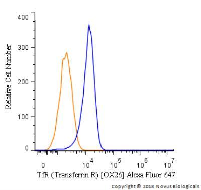
- Flow Cytometry: TfR (Transferrin R) Antibody (OX26) [NB200-585] - An intracellular stain was performed on PC12 cells with TfR (Transferrin R) [OX26] Antibody NB200-585AF647 (blue) and a matched isotype control (orange). Cells were fixed with 4% PFA and then permeabilized with 0.1% saponin. Cells were incubated in an antibody dilution of 2.5 ug/mL for 30 minutes at room temperature. Both antibodies were conjugated to Alexa Fluor 647.
- Submitted by
- Novus Biologicals (provider)
- Main image

- Experimental details
- Flow Cytometry: TfR (Transferrin R) Antibody (OX26) [NB200-585] - Analysis using the Biotin conjugate of NB200-585. Staining of mouse spleen.