Explore
 Validate
 Learn
Learn Western blot
 Immunocytochemistry
Antibody data
- Antibody Data
- Antigen structure
- References [1]
- Comments [0]
- Validations
- Immunocytochemistry [1]
- Other assay [1]
Submit
Validation data
Reference
Comment
Report error
- Product number
- MA1-21562 - Provider product page

- Provider
- Invitrogen Antibodies
- Product name
- Transferrin Receptor Monoclonal Antibody (MEM-189)
- Antibody type
- Monoclonal
- Antigen
- Other
- Description
- Store product as a concentrated solution. Centrifuge briefly prior to opening the vial.
- Reactivity
- Human
- Host
- Mouse
- Isotype
- IgG
- Antibody clone number
- MEM-189
- Vial size
- 100 µg
- Concentration
- 1 mg/mL
- Storage
- 4° C, do not freeze
Submitted references A perfused human blood-brain barrier on-a-chip for high-throughput assessment of barrier function and antibody transport.
Wevers NR, Kasi DG, Gray T, Wilschut KJ, Smith B, van Vught R, Shimizu F, Sano Y, Kanda T, Marsh G, Trietsch SJ, Vulto P, Lanz HL, Obermeier B
Fluids and barriers of the CNS 2018 Aug 31;15(1):23
Fluids and barriers of the CNS 2018 Aug 31;15(1):23
No comments: Submit comment
Supportive validation
- Submitted by
- Invitrogen Antibodies (provider)
- Main image
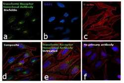
- Experimental details
- Immunofluorescence analysis of Transferrin Receptor was performed using 70% confluent log phase HeLa cells treated with BrefeldinA (5 µg/mL for 30 minutes). The cells were fixed with 4% paraformaldehyde for 10 minutes, permeabilized with 0.1% Triton™ X-100 for 15 minutes, and blocked with 1% BSA for 1 hour at room temperature. The cells were labeled with Transferrin Receptor Monoclonal Antibody (Product # MA1-21562) at 1:100 dilution in 0.1% BSA, incubated at 4 degree Celsius overnight and then labeled with Goat anti-Mouse IgG (H+L) Superclonal™ Secondary Antibody, Alexa Fluor® 488 conjugate (Product # A28175) at a dilution of 1:2000 for 45 minutes at room temperature (Panel a: green). Nuclei (Panel b: blue) were stained with ProLong™ Diamond Antifade Mountant with DAPI (Product # P36962). F-actin (Panel c: red) was stained with Rhodamine Phalloidin (Product # R415, 1:300). Panel d represents the merged image showing increased accumulation in the golgi network upon BrefeldinA treatment. Panel e represents the control cells showing cytoplasmic staining. Panel f represents control cells with no primary antibody to assess background. The images were captured at 60X magnification.
Supportive validation
- Submitted by
- Invitrogen Antibodies (provider)
- Main image

- Experimental details
- Additional file 4. Characterization of the human transferrin receptor in TY10 endothelial cells. ( a ) Immunofluorescent staining of the hTfR in TY10 endothelial cells. Scale bar is 50 um. ( b ) Flow cytometry analysis of cell surface binding of anti-TfR MEM-189 to TY10 endothelial cells in the presence and absence of transferrin (25 ug/mL), EC 50 = 0.44 +- 0.09 nM (-Tf); 0.5 +- 0.1 nM (+Tf).