Explore
 Validate
 Learn
Learn Western blot
 Immunocytochemistry
Antibody data
- Antibody Data
- Antigen structure
- References [2]
- Comments [0]
- Validations
- Western blot [1]
Submit
Validation data
Reference
Comment
Report error
- Product number
- PB9233 - Provider product page

- Provider
- Boster Biological Technology
- Product name
- Anti-Transferrin Receptor/TFRC Antibody Picoband™
- Antibody type
- Polyclonal
- Description
- Polyclonal antibody for TRANSFERRIN RECEPTOR/TFRC detection. Host: Rabbit.Size: 100μg/vial. Tested applications: WB, IHC-P, IHC-F, ICC/IF, FCM. Reactive species: Human. TRANSFERRIN RECEPTOR/TFRC information: Molecular Weight: 84871 MW; Subcellular Localization: Cell membrane ; Single-pass type II membrane protein . Melanosome . Identified by mass spectrometry in melanosome fractions from stage I to stage IV.
- Reactivity
- Human
- Host
- Rabbit
- Vial size
- 100μg/vial
- Concentration
- Add 0.2ml of distilled water will yield a concentration of 500ug/ml.
- Storage
- At -20°C for one year. After reconstitution, at 4°C for one month. It can also be aliquoted and stored frozen at -20°C for a longer time. Avoid repeated freezing and thawing.
- Handling
- Add 0.2ml of distilled water will yield a concentration of 500ug/ml.
Submitted references Novel insights into immune-related genes associated with type 2 diabetes mellitus-related cognitive impairment.
Sulfasalazine‑induced ferroptosis in breast cancer cells is reduced by the inhibitory effect of estrogen receptor on the transferrin receptor.
Gao J, Zou Y, Lv XY, Chen L, Hou XG
World journal of diabetes 2024 Apr 15;15(4):735-757
World journal of diabetes 2024 Apr 15;15(4):735-757
Sulfasalazine‑induced ferroptosis in breast cancer cells is reduced by the inhibitory effect of estrogen receptor on the transferrin receptor.
Yu H, Yang C, Jian L, Guo S, Chen R, Li K, Qu F, Tao K, Fu Y, Luo F, Liu S
Oncology reports 2019 Aug;42(2):826-838
Oncology reports 2019 Aug;42(2):826-838
No comments: Submit comment
Supportive validation
- Submitted by
- Boster Biological Technology (provider)
- Main image

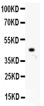
- Experimental details
- Western blot analysis of TFRC using anti-TFRC antibody (PB9233). Electrophoresis was performed on a 5-20% SDS-PAGE gel at 70V (Stacking gel) / 90V (Resolving gel) for 2-3 hours. Lane 1: Recombinant Human TFRC Protein 0.5ng After Electrophoresis, proteins were transferred to a Nitrocellulose membrane at 150mA for 50-90 minutes. Blocked the membrane with 5% Non-fat Milk/ TBS for 1.5 hour at RT. The membrane was incubated with rabbit anti-TFRC antigen affinity purified polyclonal antibody (Catalog # PB9233) at 0.5 μg/mL overnight at 4°C, then washed with TBS-0.1%Tween 3 times with 5 minutes each and probed with a goat anti-rabbit IgG-HRP secondary antibody at a dilution of 1:10000 for 1.5 hour at RT. The signal is developed using an Enhanced Chemiluminescent detection (ECL) kit (Catalog # EK1002) with Tanon 5200 system. A specific band was detected for TFRC at approximately 45KD. The expected band size for TFRC is at 45KD.
- Additional image