 Explore
 Validate
 Learn
Learn Western blot
 Immunocytochemistry
Antibody data
- Antibody Data
- Antigen structure
- References [0]
- Comments [0]
- Validations
- Immunocytochemistry [1]
- Flow cytometry [1]
Submit
Validation data
Reference
Comment
Report error
- Product number
- MA1-19299 - Provider product page

- Provider
- Invitrogen Antibodies
- Product name
- Transferrin Receptor Monoclonal Antibody (MEM-189)
- Antibody type
- Monoclonal
- Antigen
- Other
- Description
- This antibody reacts with an extracellular epitope on CD71 antigen (transferrin receptor), a 95 kDa type II homodimeric transmembrane glycoprotein expressed on activated B and lymphocytes, macrophages and erythroid precursors; it is lost on resting blood leukocytes. The antibody MEM-189 does not block binding of transferrin to the receptor. Western blot should be performed under non-reducing conditions.
- Reactivity
- Human
- Host
- Mouse
- Isotype
- IgG
- Antibody clone number
- MEM-189
- Vial size
- 100 µg
- Concentration
- 1 mg/mL
- Storage
- 4° C, do not freeze
No comments: Submit comment
Supportive validation
- Submitted by
- Invitrogen Antibodies (provider)
- Main image
- Experimental details
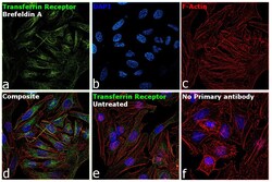
- Immunofluorescence analysis of Transferrin Receptor was performed using 70% confluent log phase HeLa cells treated with Brefeldin A (5 µg/mL for 30 minutes). The cells were fixed with 4% paraformaldehyde for 10 minutes, permeabilized with 0.1% Triton™ X-100 for 15 minutes, and blocked with 2% BSA for 1 hour at room temperature. The cells were labeled with Transferrin Receptor Monoclonal Antibody (MEM-189) (Product # MA1-19299) at 1:100 dilution in 0.1% BSA, incubated at 4 degree Celsius overnight and then labeled with Goat anti-Mouse IgG (H+L), Superclonal™ Recombinant Secondary Antibody, Alexa Fluor 488 conjugate (Product # A28175) at a dilution of 1:2000 for 45 minutes at room temperature (Panel a: green). Nuclei (Panel b: blue) were stained with SlowFade® Gold Antifade Mountant with DAPI (Product # S36938). F-actin (Panel c: red) was stained with Rhodamine Phalloidin (Product # R415, 1:300). Panel d represents the merged image showing increased Transferrin Receptor expression and localization to golgi and cytoplasm upon treatment with Brefeldin A. Panel e shows untreated cells with lower expression of Transferrin Receptor. Panel f represents control cells with no primary antibody to assess background. The images were captured at 60X magnification.

Supportive validation
- Submitted by
- Invitrogen Antibodies (provider)
- Main image
- Experimental details
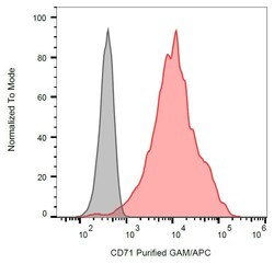
- Separation of K562 cells stained using anti-human CD71 (MEM-189) purified Monoclonal antibody (Product # MA1-19299) (concentration in sample 4 µg/mL, GAM APC, red) from K562 cells unstained by primary antibody (GAM APC, grey) in flow cytometry analysis (surface staining).
