Explore
 Validate
 Learn
Learn Western blot
Antibody data
- Antibody Data
- Antigen structure
- References [0]
- Comments [0]
- Validations
- Western blot [2]
- Immunocytochemistry [1]
Submit
Validation data
Reference
Comment
Report error
- Product number
- MA5-24106 - Provider product page

- Provider
- Invitrogen Antibodies
- Product name
- S100P Monoclonal Antibody (357517)
- Antibody type
- Monoclonal
- Antigen
- Recombinant full-length protein
- Description
- In direct ELISAs and Western blots, no cross-reactivity with recombinant human (rh) S100A10 or rhS100B is observed. Reconstitute at 0.5 mg/mL in sterile PBS.
- Reactivity
- Human
- Host
- Mouse
- Isotype
- IgG
- Antibody clone number
- 357517
- Vial size
- 100 µg
- Concentration
- 0.5 mg/mL
- Storage
- -20° C, Avoid Freeze/Thaw Cycles
No comments: Submit comment
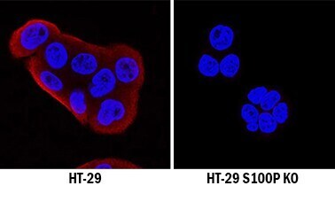
Supportive validation
- Submitted by
- Invitrogen Antibodies (provider)
- Main image

- Experimental details
- Knockout validation by Western blot analysis of S100P in immersion fixed HT‚29 human colon adenocarcinoma cell line and S100P knockout (KO) HT‚29 Human cell line. Samples were incubated in S100P monoclonal antibody (Product # MA5-24106) using a dilution of 0.1 µg/mL for 3 hours at room temperature followed by a NorthernLights™ 557-conjugated Anti-Goat IgG secondary antibody. S100P was detected in the fixed HT‚29 human colon adenocarcinoma cell line but is not detected in the knockout (KO) HT‚29 Human cell line. Cells were counterstained with DAPI (blue). Specific staining was localized to cytoplasm. View our protocol for Fluorescent ICC Staining of Cells on Coverslips.
- Submitted by
- Invitrogen Antibodies (provider)
- Main image
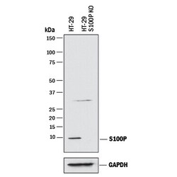
- Experimental details
- Western blot analysis of S100P in HT‚29 human colon adenocarcinoma parental cell line and S100P knockout HT‚29 cell line (KO). Samples were incubated in S100P monoclonal antibody (Product # MA5-24106) using a dilution of 1 µg/mL followed by a HRP-conjugated Anti-Mouse IgG secondary antibody. A specific band was detected for S100P at approximately 10 kDa (as indicated) in the parental HT‚29cell line, but is not detectable in knockout HT‚29 cell line. GAPDH is shown as a loading control. This experiment was conducted under reducing conditions.

Supportive validation
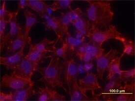
- Submitted by
- Invitrogen Antibodies (provider)
- Main image

- Experimental details
- Immunocytochemistry analysis of S100P in immersion fixed NTera-2 human testicular embryonic carcinoma cell line. Samples were incubated in S100P monoclonal antibody (Product # MA5-24106) using a dilution of 10 µg/mL for 3 hours at room temperature followed by NorthernLights™ 557-conjugated Anti-Mouse IgG Secondary Antibody (red) and counterstained with DAPI (blue).