Explore
 Validate
 Learn
Learn Western blot
Antibody data
- Antibody Data
- Antigen structure
- References [5]
- Comments [0]
- Validations
- Western blot [1]
- Immunocytochemistry [2]
Submit
Validation data
Reference
Comment
Report error
- Product number
- MAB2957 - Provider product page

- Provider
- R&D Systems
- Product name
- Human S100P Antibody
- Antibody type
- Monoclonal
- Description
- Protein A or G purified from hybridoma culture supernatant. Detects human S100P in direct ELISAs and Western blots. In direct ELISAs and Western blots, no cross-reactivity with recombinant human (rh) S100A10 or rhS100B is observed.
- Reactivity
- Human
- Host
- Mouse
- Conjugate
- Unconjugated
- Antigen sequence
P25815- Isotype
- IgG
- Antibody clone number
- 357517
- Vial size
- 100 ug
- Concentration
- LYOPH
- Storage
- Use a manual defrost freezer and avoid repeated freeze-thaw cycles. 12 months from date of receipt, -20 to -70 °C as supplied. 1 month, 2 to 8 °C under sterile conditions after reconstitution. 6 months, -20 to -70 °C under sterile conditions after reconstitution.
Submitted references Dichotomy in intrahepatic cholangiocarcinomas based on histologic similarities to hilar cholangiocarcinomas.
Novel quantitative analysis of the S100P protein combined with endoscopic ultrasound-guided fine needle aspiration cytology in the diagnosis of pancreatic adenocarcinoma.
S100P expression in response to sex steroids during the implantation window in human endometrium.
S100P-derived RAGE antagonistic peptide reduces tumor growth and metastasis.
Morphoproteomic profile of mTOR, Ras/Raf kinase/ERK, and NF-kappaB pathways in human gastric adenocarcinoma.
Akita M, Fujikura K, Ajiki T, Fukumoto T, Otani K, Azuma T, Itoh T, Ku Y, Zen Y
Modern pathology : an official journal of the United States and Canadian Academy of Pathology, Inc 2017 Jul;30(7):986-997
Modern pathology : an official journal of the United States and Canadian Academy of Pathology, Inc 2017 Jul;30(7):986-997
Novel quantitative analysis of the S100P protein combined with endoscopic ultrasound-guided fine needle aspiration cytology in the diagnosis of pancreatic adenocarcinoma.
Chiba M, Imazu H, Kato M, Ikeda K, Arakawa H, Kato T, Sumiyama K, Homma S
Oncology reports 2017 Apr;37(4):1943-1952
Oncology reports 2017 Apr;37(4):1943-1952
S100P expression in response to sex steroids during the implantation window in human endometrium.
Zhang D, Ma C, Sun X, Xia H, Zhang W
Reproductive biology and endocrinology : RB&E 2012 Dec 7;10:106
Reproductive biology and endocrinology : RB&E 2012 Dec 7;10:106
S100P-derived RAGE antagonistic peptide reduces tumor growth and metastasis.
Arumugam T, Ramachandran V, Gomez SB, Schmidt AM, Logsdon CD
Clinical cancer research : an official journal of the American Association for Cancer Research 2012 Aug 15;18(16):4356-64
Clinical cancer research : an official journal of the American Association for Cancer Research 2012 Aug 15;18(16):4356-64
Morphoproteomic profile of mTOR, Ras/Raf kinase/ERK, and NF-kappaB pathways in human gastric adenocarcinoma.
Feng W, Brown RE, Trung CD, Li W, Wang L, Khoury T, Alrawi S, Yao J, Xia K, Tan D
Annals of clinical and laboratory science 2008 Summer;38(3):195-209
Annals of clinical and laboratory science 2008 Summer;38(3):195-209
No comments: Submit comment
Supportive validation
- Submitted by
- R&D Systems (provider)
- Main image
- Experimental details
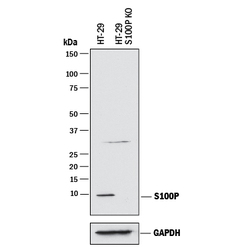
- Detection of Human S100P by Western Blot. Western blot shows lysates of HT-29 human colon adenocarcinoma parental cell line and S100P knockout HT-29 cell line (KO). PVDF membrane was probed with 1 µg/mL of Mouse Anti-Human S100P Monoclonal Antibody (Catalog # MAB2957) followed by HRP-conjugated Anti-Mouse IgG Secondary Antibody (Catalog # HAF018). A specific band was detected for S100P at approximately 10 kDa (as indicated) in the parental HT-29 cell line, but is not detectable in knockout HT-29 cell line. GAPDH (Catalog # AF5718) is shown as a loading control. This experiment was conducted under reducing conditions and using Immunoblot Buffer Group 1.

Supportive validation
- Submitted by
- R&D Systems (provider)
- Main image
- Experimental details
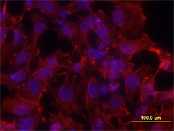
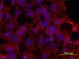
- S100P in NTera-2 Human Cell Line. S100P was detected in immersion fixed NTera-2 human testicular embryonic carcinoma cell line using 10 µg/mL Mouse Anti-Human S100P Monoclonal Antibody (Catalog # MAB2957) for 3 hours at room temperature. Cells were stained with the NorthernLights™ 557-conjugated Anti-Mouse IgG Secondary Antibody (red; Catalog # NL007) and counterstained with DAPI (blue). View our protocol for Fluorescent ICC Staining of Cells on Coverslips.
- Submitted by
- R&D Systems (provider)
- Main image

- Experimental details
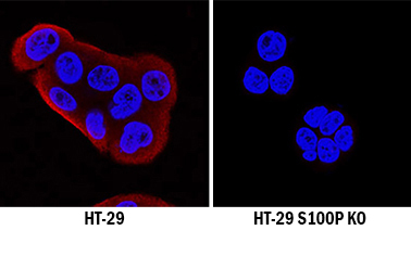
- S100P Specificity is Shown by Immunocytochemistry in Knockout Cell Line. S100P was detected in immersion fixed HT-29 human colon adenocarcinoma cell line but is not detected in S100P knockout (KO) HT-29 Human Cell Line cell line using Mouse Anti-Human S100P Monoclonal Antibody (Catalog # MAB2957) at 0.1 µg/mL for 3 hours at room temperature. Cells were stained using the NorthernLights 557-conjugated Anti-Goat IgG Secondary Antibody (red; Catalog # NL001) and counterstained with DAPI (blue). Specific staining was localized to cytoplasm. View our protocol for Fluorescent ICC Staining of Cells on Coverslips.