 Explore
 Validate
 Learn
Learn Western blot
 ELISA
ELISAAntibody data
- Antibody Data
- Antigen structure
- References [9]
- Comments [0]
- Validations
- Western blot [1]
- Immunocytochemistry [1]
- Immunohistochemistry [2]
Submit
Validation data
Reference
Comment
Report error
- Product number
- 21749-1-AP - Provider product page

- Provider
- Proteintech Group
- Proper citation
- Proteintech Cat#21749-1-AP, RRID:AB_10858789
- Product name
- HIGD1A antibody
- Antibody type
- Polyclonal
- Description
- KD/KO validated HIGD1A antibody (Cat. #21749-1-AP) is a rabbit polyclonal antibody that shows reactivity with human, rat, mouse and has been validated for the following applications: IF, IHC, WB,ELISA.
- Reactivity
- Human, Mouse, Rat
- Host
- Rabbit
- Conjugate
- Unconjugated
- Isotype
- IgG
- Vial size
- 20ul, 150ul
Submitted references Affinity-Based Protein Profiling Revealed that HIGD1A is a Direct Target Protein of Aristolochic Acids.
Elevated expression of HIGD1A drives hepatocellular carcinoma progression by regulating polyamine metabolism through c-Myc-ODC1 nexus.
HIGD1A links SIRT1 activity to adipose browning by inhibiting the ROS/DNA damage pathway.
DNA damage-induced translocation of mitochondrial factor HIGD1A into the nucleus regulates homologous recombination and radio/chemo-sensitivity.
HIG1 domain family member 1A disrupts proliferation, migration, and invasion of colon adenocarcinoma cells.
HIGD2A is Required for Assembly of the COX3 Module of Human Mitochondrial Complex IV.
Higd1a improves respiratory function in the models of mitochondrial disorder.
Higd1a is a positive regulator of cytochrome c oxidase.
Nuclear localization of the mitochondrial factor HIGD1A during metabolic stress.
Gong Y, Zhang S, Wei A, Ji X, Chong X, Cen J, Zhao X, Luo Z, Pei Z, Mao G, Zhang X, Sun M, Sun Z, Yin Z, Zou Z, Meng WQ
Advanced science (Weinheim, Baden-Wurttemberg, Germany) 2026 Jan;13(5):e13117
Advanced science (Weinheim, Baden-Wurttemberg, Germany) 2026 Jan;13(5):e13117
Elevated expression of HIGD1A drives hepatocellular carcinoma progression by regulating polyamine metabolism through c-Myc-ODC1 nexus.
Zhang H, Li X, Liu Z, Lin Z, Huang K, Wang Y, Chen Y, Liao L, Wu L, Xie Z, Hou J, Zhang X, Liu H
Cancer & metabolism 2024 Feb 23;12(1):7
Cancer & metabolism 2024 Feb 23;12(1):7
HIGD1A links SIRT1 activity to adipose browning by inhibiting the ROS/DNA damage pathway.
Li BY, Peng WQ, Liu Y, Guo L, Tang QQ
Cell reports 2023 Jul 25;42(7):112731
Cell reports 2023 Jul 25;42(7):112731
DNA damage-induced translocation of mitochondrial factor HIGD1A into the nucleus regulates homologous recombination and radio/chemo-sensitivity.
Chen B, Xu F, Gao Y, Hu G, Zhu K, Lu H, Xu A, Chen S, Wu L, Zhao G
Oncogene 2022 Mar;41(13):1918-1930
Oncogene 2022 Mar;41(13):1918-1930
HIG1 domain family member 1A disrupts proliferation, migration, and invasion of colon adenocarcinoma cells.
Xu Z, Sun J, Mao Y, Chen Y, Zhang T, Qin Y, Hua D
Bioengineered 2021 Dec;12(2):10501-10511
Bioengineered 2021 Dec;12(2):10501-10511
HIGD2A is Required for Assembly of the COX3 Module of Human Mitochondrial Complex IV.
Hock DH, Reljic B, Ang CS, Muellner-Wong L, Mountford HS, Compton AG, Ryan MT, Thorburn DR, Stroud DA
Molecular & cellular proteomics : MCP 2020 Jul;19(7):1145-1160
Molecular & cellular proteomics : MCP 2020 Jul;19(7):1145-1160
Higd1a improves respiratory function in the models of mitochondrial disorder.
Nagao T, Shintani Y, Hayashi T, Kioka H, Kato H, Nishida Y, Yamazaki S, Tsukamoto O, Yashirogi S, Yazawa I, Asano Y, Shinzawa-Itoh K, Imamura H, Suzuki T, Suzuki T, Goto YI, Takashima S
FASEB journal : official publication of the Federation of American Societies for Experimental Biology 2020 Jan;34(1):1859-1871
FASEB journal : official publication of the Federation of American Societies for Experimental Biology 2020 Jan;34(1):1859-1871
Higd1a is a positive regulator of cytochrome c oxidase.
Hayashi T, Asano Y, Shintani Y, Aoyama H, Kioka H, Tsukamoto O, Hikita M, Shinzawa-Itoh K, Takafuji K, Higo S, Kato H, Yamazaki S, Matsuoka K, Nakano A, Asanuma H, Asakura M, Minamino T, Goto Y, Ogura T, Kitakaze M, Komuro I, Sakata Y, Tsukihara T, Yoshikawa S, Takashima S
Proceedings of the National Academy of Sciences of the United States of America 2015 Feb 3;112(5):1553-8
Proceedings of the National Academy of Sciences of the United States of America 2015 Feb 3;112(5):1553-8
Nuclear localization of the mitochondrial factor HIGD1A during metabolic stress.
Ameri K, Rajah AM, Nguyen V, Sanders TA, Jahangiri A, Delay M, Donne M, Choi HJ, Tormos KV, Yeghiazarians Y, Jeffrey SS, Rinaudo PF, Rowitch DH, Aghi M, Maltepe E
PloS one 2013;8(4):e62758
PloS one 2013;8(4):e62758
No comments: Submit comment
Supportive validation
- Submitted by
- Proteintech Group (provider)
- Main image

- Experimental details
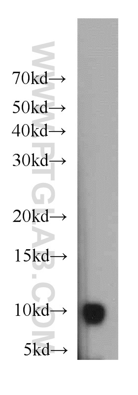
- HeLa cells were subjected to SDS PAGE followed by western blot with 21749-1-AP(HIGD1A antibody) at dilution of 1:500
- Sample type
- cell line
Supportive validation
- Submitted by
- Proteintech Group (provider)
- Main image
- Experimental details
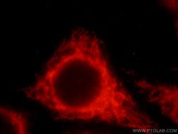
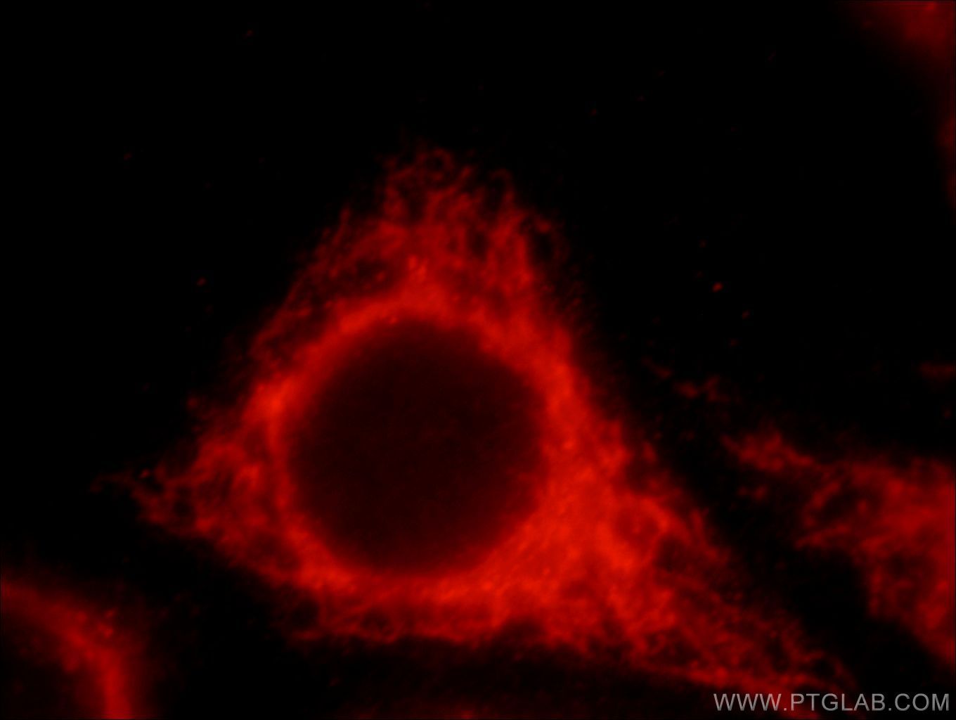
- Immunofluorescent analysis of HepG2 cells, using HIGD1A antibody 21749-1-AP at 1:25 dilution and Rhodamine-labeled goat anti-rabbit IgG (red).
- Sample type
- cell line
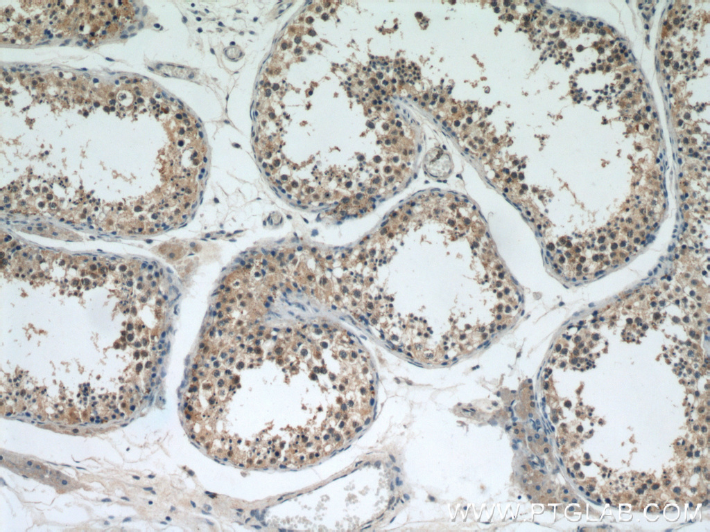
Supportive validation
- Submitted by
- Proteintech Group (provider)
- Main image
- Experimental details
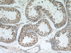
- Immunohistochemical of paraffin-embedded human testis using 21749-1-AP(HIGD1A antibody) at dilution of 1:200 (under 10x lens)
- Sample type
- tissue
- Submitted by
- Proteintech Group (provider)
- Main image
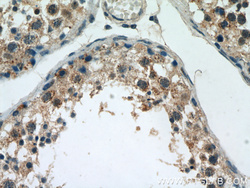
- Experimental details
- Immunohistochemical of paraffin-embedded human testis using 21749-1-AP(HIGD1A antibody) at dilution of 1:200 (under 40x lens)
- Sample type
- tissue
