Explore
 Validate
 Learn
LearnMA5-15928
antibody from Invitrogen Antibodies
Targeting: SKP1
EMC19, MGC34403, OCP-II, OCP2, p19A, SKP1A, TCEB1L
 Western blot
 ELISA
ELISA Immunocytochemistry
Antibody data
- Antibody Data
- Antigen structure
- References [2]
- Comments [0]
- Validations
- Immunocytochemistry [4]
- Immunohistochemistry [1]
- Flow cytometry [2]
Submit
Validation data
Reference
Comment
Report error
- Product number
- MA5-15928 - Provider product page

- Provider
- Invitrogen Antibodies
- Product name
- SKP1 Monoclonal Antibody (4E11)
- Antibody type
- Monoclonal
- Antigen
- Purifed from natural sources
- Description
- MA5-15928 targets SKP1 in indirect ELISA, FACS, IF, IHC, and WB applications and shows reactivity with Human samples. The MA5-15928 immunogen is purified recombinant fragment of human SKP1 expressed in E. Coli. . MA5-15928 detects SKP1 which has a predicted molecular weight of approximately 19kDa.
- Reactivity
- Human, Mouse
- Host
- Mouse
- Isotype
- IgG
- Antibody clone number
- 4E11
- Vial size
- 100 μL
- Concentration
- Conc. Not Determined
- Storage
- Store at 4°C short term. For long term storage, store at -20°C, avoiding freeze/thaw cycles.
Submitted references Differential sensitivity of acute myeloid leukemia cells to daunorubicin depends on P2X7A versus P2X7B receptor expression.
Cand1-Mediated Adaptive Exchange Mechanism Enables Variation in F-Box Protein Expression.
Pegoraro A, Orioli E, De Marchi E, Salvestrini V, Milani A, Di Virgilio F, Curti A, Adinolfi E
Cell death & disease 2020 Oct 18;11(10):876
Cell death & disease 2020 Oct 18;11(10):876
Cand1-Mediated Adaptive Exchange Mechanism Enables Variation in F-Box Protein Expression.
Liu X, Reitsma JM, Mamrosh JL, Zhang Y, Straube R, Deshaies RJ
Molecular cell 2018 Mar 1;69(5):773-786.e6
Molecular cell 2018 Mar 1;69(5):773-786.e6
No comments: Submit comment
Supportive validation
- Submitted by
- Invitrogen Antibodies (provider)
- Main image

- Experimental details
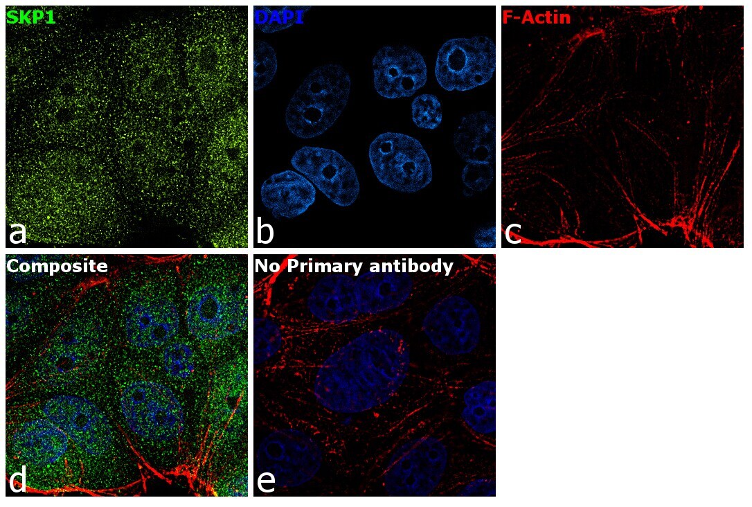
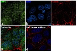
- Immunofluorescence analysis of SKP1 was performed using 70% confluent log phase MCF-7 cells. The cells were fixed with 4% paraformaldehyde for 10 minutes, permeabilized with 0.1% Triton™ X-100 for 15 minutes, and blocked with 1% BSA for 1 hour at room temperature. The cells were labeled with SKP1 Mouse Monoclonal Antibody (4E11) (Product # MA5-15928) at 1:100 dilution in 0.1% BSA, incubated at 4 degree Celsius overnight and then labeled with Goat anti-Mouse IgG (H+L) Superclonal™ Secondary Antibody, Alexa Fluor® 488 conjugate (Product # A28175) at a dilution of 1:2000 for 45 minutes at room temperature (Panel a: green). Nuclei (Panel b: blue) were stained with SlowFade® Gold Antifade Mountant with DAPI (Product # S36938). F-actin (Panel c: red) was stained with Rhodamine Phalloidin (Product # R415, 1:300). Panel d represents the merged image showing cytoplasmic and nuclear localization. Panel e represents control cells with no primary antibody to assess background. The images were captured at 60X magnification.
- Submitted by
- Invitrogen Antibodies (provider)
- Main image

- Experimental details
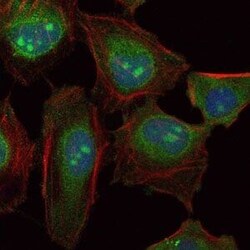
- Immunofluorescence analysis of HeLa cells using SKP1 monoclonal antibody (Product # MA5-15928) (Green). Blue: DRAQ5 fluorescent DNA dye. Red: actin filaments have been labeled with phalloidin.

- Submitted by
- Invitrogen Antibodies (provider)
- Main image
- Experimental details
- Immunofluorescence analysis of HeLa cells using SKP1 monoclonal antibody (Product # MA5-15928) (Green). Blue: DRAQ5 fluorescent DNA dye. Red: actin filaments have been labeled with phalloidin.

- Submitted by
- Invitrogen Antibodies (provider)
- Main image
- Experimental details
- Immunofluorescence analysis of SKP1 was performed using 70% confluent log phase MCF-7 cells. The cells were fixed with 4% paraformaldehyde for 10 minutes, permeabilized with 0.1% Triton™ X-100 for 15 minutes, and blocked with 1% BSA for 1 hour at room temperature. The cells were labeled with SKP1 Mouse Monoclonal Antibody (4E11) (Product # MA5-15928) at 1:100 dilution in 0.1% BSA, incubated at 4 degree Celsius overnight and then labeled with Goat anti-Mouse IgG (H+L) Superclonal™ Secondary Antibody, Alexa Fluor® 488 conjugate (Product # A28175) at a dilution of 1:2000 for 45 minutes at room temperature (Panel a: green). Nuclei (Panel b: blue) were stained with SlowFade® Gold Antifade Mountant with DAPI (Product # S36938). F-actin (Panel c: red) was stained with Rhodamine Phalloidin (Product # R415, 1:300). Panel d represents the merged image showing cytoplasmic and nuclear localization. Panel e represents control cells with no primary antibody to assess background. The images were captured at 60X magnification.

Supportive validation
- Submitted by
- Invitrogen Antibodies (provider)
- Main image

- Experimental details
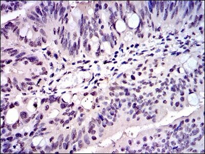
- Immunohistochemical analysis of paraffin-embedded rectum cancer tissues using SKP1 monoclonal antibody (Product # MA5-15928) followed with DAB staining.
Supportive validation
- Submitted by
- Invitrogen Antibodies (provider)
- Main image

- Experimental details
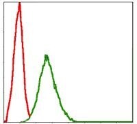
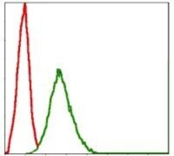
- Flow cytometric analysis of NIH/3T3 cells using SKP1 monoclonal antibody (Product # MA5-15928) (green) and negative control (red).

- Submitted by
- Invitrogen Antibodies (provider)
- Main image
- Experimental details
- Flow cytometric analysis of NIH/3T3 cells using SKP1 monoclonal antibody (Product # MA5-15928) (green) and negative control (red).