Explore
 Validate
 Learn
Learn Western blot
Antibody data
- Antibody Data
- Antigen structure
- References [0]
- Comments [0]
- Validations
- Western blot [1]
- Immunocytochemistry [1]
- Immunohistochemistry [1]
- Flow cytometry [1]
Submit
Validation data
Reference
Comment
Report error
- Product number
- AP12394PU-N - Provider product page

- Provider
- Acris Antibodies GmbH
- Proper citation
- Acris Antibodies GmbH Cat#AP12394PU-N, RRID:AB_1752635
- Product name
- anti FZD1 / Frizzled-1 (Center)
- Antibody type
- Polyclonal
- Antigen
- KLH conjugated synthetic peptide between 374~404 amino acids from the center region of Human FZD1.
- Reactivity
- Human
- Host
- Rabbit
- Vial size
- 0.4 ml
- Concentration
- 2.0 mg/mL
No comments: Submit comment
Supportive validation
- Submitted by
- Acris Antibodies GmbH (provider)
- Main image
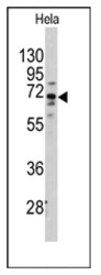
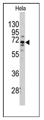
- Experimental details
- Western blot analysis of anti-FZD1 (Center) (Cat.#AP12394PU-N) in Hela cell line lysates (35ug/lane). FZD1 (arrow) was detected using the purified Pab (1:60 dilution).
Supportive validation
- Submitted by
- Acris Antibodies GmbH (provider)
- Main image
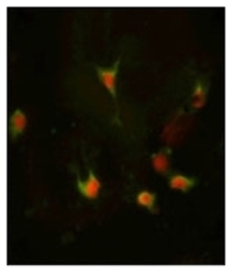
- Experimental details
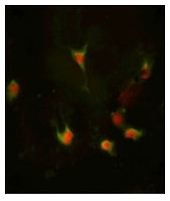
- Immunofluorescence analysis of FZD1 Antibody (Center) with hela cells. 0.025 mg/ml primary antibody was followed by FITC-conjugated Goat anti-Rabbit IgG (whole molecule). FITC emits green fluorescence.Red counterstaining is PI.
Supportive validation
- Submitted by
- Acris Antibodies GmbH (provider)
- Main image
- Experimental details
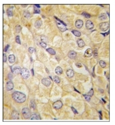
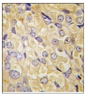
- Formalin-fixed and paraffin-embedded human prostata carcinoma tissue reacted with FZD1 antibody (Center) (Cat.#AP12394PU-N), which was peroxidase-conjugated to the secondary antibody, followed by DAB staining. This data demonstrates the use of this antibody for immunohistochemistry; clinical relevance has not been evaluated.
Supportive validation
- Submitted by
- Acris Antibodies GmbH (provider)
- Main image

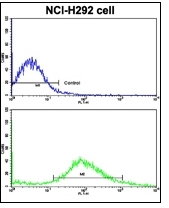
- Experimental details
- Flow Cytometric analysis of NCI-H292 cells using FZD1 Antibody (Center)(bottom Histogram) compared to a Negative Control cell (top histogram). FITC-conjugated Goat-anti-Rabbit secondary antibodies were used for the analysis.