 Explore
 Validate
 Learn
Learn Western blot
Antibody data
- Antibody Data
- Antigen structure
- References [1]
- Comments [0]
- Validations
- Western blot [1]
- Immunohistochemistry [1]
Submit
Validation data
Reference
Comment
Report error
- Product number
- MAB701 - Provider product page

- Provider
- Novus Biologicals
- Product name
- Mouse Monoclonal Ubiquitin/Ubiquitin+1 Antibody
- Antibody type
- Monoclonal
- Description
- Protein A or G purified from hybridoma culture supernatant. Detects human Ubiquitin/Ubiquitin+1 in Western blots..
- Reactivity
- Human
- Host
- Mouse
- Isotype
- IgG
- Vial size
- 100 ug
- Concentration
- LYOPH
- Storage
- Use a manual defrost freezer and avoid repeated freeze-thaw cycles. 12 months from date of receipt, -20 to -70 degreesC as supplied. 1 month, 2 to 8 degreesC under sterile conditions after reconstitution. 6 months, -20 to -70 degreesC under sterile conditions after reconstitution.
Submitted references Ubiquitin as potential cerebrospinal fluid marker of Creutzfeldt-Jakob disease.
Steinacker P, Rist W, Swiatek-de-Lange M, Lehnert S, Jesse S, Pabst A, Tumani H, von Arnim CA, Mitrova E, Kretzschmar HA, Lenter M, Wiltfang J, Otto M
Proteomics 2010 Jan;10(1):81-9
Proteomics 2010 Jan;10(1):81-9
No comments: Submit comment
Supportive validation
- Submitted by
- Novus Biologicals (provider)
- Main image
- Experimental details
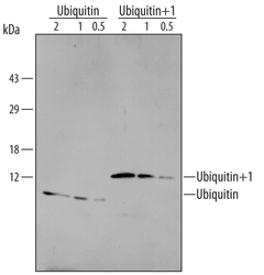
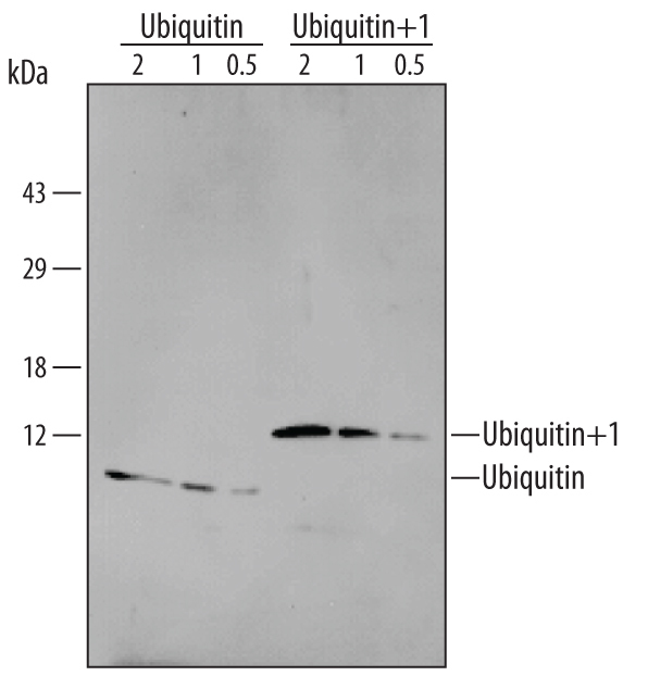
- Detection of Human Ubiquitin/Ubiquitin+1 by Western Blot. Western blot shows samples of Recombinant Human Ubiquitin (Catalog # 701-UB) (2, 1, and 0.5 ng) and Recombinant Human Ubiquitin+1 (Catalog # 703-UB) (2, 1, and 0.5 ng). PVDF membrane was probed with 1-2 µg/mL Mouse Anti-Human Ubiquitin/Ubiquitin+1 Monoclonal Antibody (Catalog # MAB701) followed by HRP-conjugated Anti-Mouse IgG Secondary Antibody (Catalog # HAF007). Specific bands for Ubiquitin and Ubiquitin+1 were detected at approximately 11 kDa and 13 kDa, respectively (as indicated). This experiment was conducted under reducing conditions and using Immunoblot Buffer Group 9.
Supportive validation
- Submitted by
- Novus Biologicals (provider)
- Main image
- Experimental details
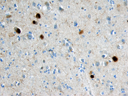
- Ubiquitin/Ubiquitin+1 in Human Alzheimer's Disease Brain. Ubiquitin/Ubiquitin+1 was detected in immersion fixed paraffin-embedded sections of human Alzheimer's disease brain (cortex) using 25 µg/mL Mouse Anti-Human Ubiquitin/ Ubiquitin+1 Monoclonal Antibody (Catalog # MAB701) overnight at 4 °C. Tissue was stained with the Anti-Mouse HRP-DAB Cell & Tissue Staining Kit (brown; Catalog # CTS002) and counter-stained with hematoxylin (blue). Specific labeling was localized to the cytoplasm of neurons in the cortex. View our protocol for Chromogenic IHC Staining of Paraffin-embedded Tissue Sections.
