Explore
 Validate
 Learn
Learn Western blot
 Flow cytometry
Antibody data
- Antibody Data
- Antigen structure
- References [5]
- Comments [0]
- Validations
- Western blot [1]
- Immunohistochemistry [1]
Submit
Validation data
Reference
Comment
Report error
- Product number
- AF1790 - Provider product page

- Provider
- Novus Biologicals
- Product name
- Goat Polyclonal Cadherin-11 Antibody
- Antibody type
- Polyclonal
- Description
- Antigen Affinity-purified. Detects human Cadherin-11 in direct ELISAs and Western blots. In direct ELISAs, less than 5% cross-reactivity with recombinant human (rh) Cadherin-7, rhCadherin-8, rhCadherin-10, rhCadherin-18 and rhCadherin-20 is observed.
- Reactivity
- Human
- Host
- Goat
- Conjugate
- Unconjugated
- Isotype
- IgG
- Vial size
- 100 ug
- Concentration
- LYOPH
- Storage
- Use a manual defrost freezer and avoid repeated freeze-thaw cycles. 12 months from date of receipt, -20 to -70 degreesC as supplied. 1 month, 2 to 8 degreesC under sterile conditions after reconstitution. 6 months, -20 to -70 degreesC under sterile conditions after reconstitution.
Submitted references N-cadherin-mediated cell-cell adhesion promotes cell migration in a three-dimensional matrix.
Cadherin-11 promotes the metastasis of prostate cancer cells to bone.
Secretion and organization of a cornea-like tissue in vitro by stem cells from human corneal stroma.
Accelerated formation of multicellular 3-D structures by cell-to-cell cross-linking.
E-cadherin cell-cell adhesion in ewing tumor cells mediates suppression of anoikis through activation of the ErbB4 tyrosine kinase.
Shih W, Yamada S
Journal of cell science 2012 Aug 1;125(Pt 15):3661-70
Journal of cell science 2012 Aug 1;125(Pt 15):3661-70
Cadherin-11 promotes the metastasis of prostate cancer cells to bone.
Chu K, Cheng CJ, Ye X, Lee YC, Zurita AJ, Chen DT, Yu-Lee LY, Zhang S, Yeh ET, Hu MC, Logothetis CJ, Lin SH
Molecular cancer research : MCR 2008 Aug;6(8):1259-67
Molecular cancer research : MCR 2008 Aug;6(8):1259-67
Secretion and organization of a cornea-like tissue in vitro by stem cells from human corneal stroma.
Du Y, Sundarraj N, Funderburgh ML, Harvey SA, Birk DE, Funderburgh JL
Investigative ophthalmology & visual science 2007 Nov;48(11):5038-45
Investigative ophthalmology & visual science 2007 Nov;48(11):5038-45
Accelerated formation of multicellular 3-D structures by cell-to-cell cross-linking.
De Bank PA, Hou Q, Warner RM, Wood IV, Ali BE, Macneil S, Kendall DA, Kellam B, Shakesheff KM, Buttery LD
Biotechnology and bioengineering 2007 Aug 15;97(6):1617-25
Biotechnology and bioengineering 2007 Aug 15;97(6):1617-25
E-cadherin cell-cell adhesion in ewing tumor cells mediates suppression of anoikis through activation of the ErbB4 tyrosine kinase.
Kang HG, Jenabi JM, Zhang J, Keshelava N, Shimada H, May WA, Ng T, Reynolds CP, Triche TJ, Sorensen PH
Cancer research 2007 Apr 1;67(7):3094-105
Cancer research 2007 Apr 1;67(7):3094-105
No comments: Submit comment
Supportive validation
- Submitted by
- Novus Biologicals (provider)
- Main image
- Experimental details
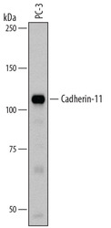
- Detection of Human Cadherin-11 by Western Blot. Western blot shows lysates of PC-3 human prostate cancer cell line. PVDF membrane was probed with 1 µg/mL of Goat Anti-Human Cadherin-11 Antigen Affinity-purified Polyclonal Antibody (Catalog # AF1790) followed by HRP-conjugated Anti-Goat IgG Secondary Antibody (Catalog # HAF109). A specific band was detected for Cadherin-11 at approximately 110 kDa (as indicated). This experiment was conducted under reducing conditions and using Immunoblot Buffer Group 1.

Supportive validation
- Submitted by
- Novus Biologicals (provider)
- Main image

- Experimental details
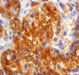
- Cadherin-11 in Human Prostate Cancer Tissue. Cadherin-11 was detected in fomralin fixed paraffin-embedded sections of human prostate cancer tissue using Goat Anti-Human Cadherin-11 Antigen Affinity-purified Polyclonal Antibody (Catalog # AF1790) at 15 µg/mL overnight at 4 °C. Tissue was stained using the Anti-Goat HRP-DAB Cell & Tissue Staining Kit (brown; Catalog # CTS008) and counterstained with hematoxylin (blue). Specific staining was localized in the membrane. View our protocol for Chromogenic IHC Staining of Paraffin-embedded Tissue Sections.