 Explore
 Validate
 Learn
Learn Flow cytometry
Antibody data
- Antibody Data
- Antigen structure
- References [3]
- Comments [0]
- Validations
- Flow cytometry [1]
Submit
Validation data
Reference
Comment
Report error
- Product number
- FAB17901G - Provider product page

- Provider
- Novus Biologicals
- Product name
- Mouse Monoclonal Cadherin-11 Antibody
- Antibody type
- Monoclonal
- Description
- Protein A or G purified from hybridoma culture supernatant. Detects human Cadherin-11 in direct ELISAs. In direct ELISAs, no cross-reactivity with recombinant human (rh) Cadherin-4, -6, -8, -12, -13, -17, rhE-Cadherin, rhN-Cadherin, rhP-Cadherin, or rhVE-Cadherin is observed.
- Reactivity
- Human
- Host
- Mouse
- Conjugate
- Green dye
- Isotype
- IgG
- Vial size
- 100 Tests
- Storage
- Protect from light. Do not freeze. 12 months from date of receipt, 2 to 8 degreesC as supplied.
Submitted references Cadherin-11 contributes to liver fibrosis induced by carbon tetrachloride.
Physiologic expansion of human heart-derived cells enhances therapeutic repair of injured myocardium.
Targeting of cadherin-11 decreases skin fibrosis in the tight skin-1 mouse model.
Pedroza M, To S, Smith J, Agarwal SK
PloS one 2019;14(7):e0218971
PloS one 2019;14(7):e0218971
Physiologic expansion of human heart-derived cells enhances therapeutic repair of injured myocardium.
Mount S, Kanda P, Parent S, Khan S, Michie C, Davila L, Chan V, Davies RA, Haddad H, Courtman D, Stewart DJ, Davis DR
Stem cell research & therapy 2019 Nov 4;10(1):316
Stem cell research & therapy 2019 Nov 4;10(1):316
Targeting of cadherin-11 decreases skin fibrosis in the tight skin-1 mouse model.
Pedroza M, Welschhans RL, Agarwal SK
PloS one 2017;12(11):e0187109
PloS one 2017;12(11):e0187109
No comments: Submit comment
Supportive validation
- Submitted by
- Novus Biologicals (provider)
- Main image
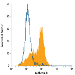
- Experimental details
- Detection of Cadherin-11 in PC-3 Human Cell Line by Flow Cytometry. PC-3 human prostate cancer cell line was stained with Mouse Anti-Human Cadherin-11 Alexa Fluor® 488-conjugated Monoclonal Antibody (Catalog # FAB17901G, filled histogram) or isotype control antibody (Catalog # IC003G, open histogram). Cells were stained in a buffer containing Ca2+ and Mg2+. View our protocol for Staining Membrane-associated Proteins.
