 Explore
 Validate
 Learn
Learn Western blot
Antibody data
- Antibody Data
- Antigen structure
- References [5]
- Comments [0]
- Validations
- Western blot [2]
- Immunohistochemistry [1]
Submit
Validation data
Reference
Comment
Report error
- Product number
- MAB1790 - Provider product page

- Provider
- R&D Systems
- Product name
- Human Cadherin-11 Antibody
- Antibody type
- Monoclonal
- Description
- Protein A or G purified from hybridoma culture supernatant. Detects human Cadherin-11 in direct ELISAs and Western blots. Does not cross-react with recombinant human (rh) Cadherin-4, rhCadherin-8, rhCadherin-12, rhCadherin-13, rhCadherin-17, rhE-Cadherin, rhN-Cadherin, rhP-Cadherin, or rhVE-Cadherin.
- Reactivity
- Human
- Host
- Mouse
- Conjugate
- Unconjugated
- Antigen sequence
AAA35622- Isotype
- IgG
- Antibody clone number
- 283416
- Vial size
- 100 ug
- Concentration
- LYOPH
- Storage
- Use a manual defrost freezer and avoid repeated freeze-thaw cycles. 12 months from date of receipt, -20 to -70 °C as supplied. 1 month, 2 to 8 °C under sterile conditions after reconstitution. 6 months, -20 to -70 °C under sterile conditions after reconstitution.
Submitted references The role of cadherin-11 in microcystin-LR-induced migration and invasion in colorectal carcinoma cells.
Cadherin-11 expression is upregulated in invasive human breast cancer.
Cadherin-11 in renal cell carcinoma bone metastasis.
N-cadherin-mediated cell-cell adhesion promotes cell migration in a three-dimensional matrix.
Hypoxia-induced abrogation of contact-dependent inhibition of rheumatoid arthritis synovial fibroblast proliferation.
Zhu Q, Wang Z, Zhou L, Ren Y, Gong Y, Qin W, Bai L, Hu J, Wang T
Oncology letters 2018 Feb;15(2):1417-1422
Oncology letters 2018 Feb;15(2):1417-1422
Cadherin-11 expression is upregulated in invasive human breast cancer.
Pohlodek K, Tan YY, Singer CF, Gschwantler-Kaulich D
Oncology letters 2016 Dec;12(6):4393-4398
Oncology letters 2016 Dec;12(6):4393-4398
Cadherin-11 in renal cell carcinoma bone metastasis.
Satcher RL, Pan T, Cheng CJ, Lee YC, Lin SC, Yu G, Li X, Hoang AG, Tamboli P, Jonasch E, Gallick GE, Lin SH
PloS one 2014;9(2):e89880
PloS one 2014;9(2):e89880
N-cadherin-mediated cell-cell adhesion promotes cell migration in a three-dimensional matrix.
Shih W, Yamada S
Journal of cell science 2012 Aug 1;125(Pt 15):3661-70
Journal of cell science 2012 Aug 1;125(Pt 15):3661-70
Hypoxia-induced abrogation of contact-dependent inhibition of rheumatoid arthritis synovial fibroblast proliferation.
Nonomura Y, Mizoguchi F, Suzuki A, Nanki T, Kato H, Miyasaka N, Kohsaka H
The Journal of rheumatology 2009 Apr;36(4):698-705
The Journal of rheumatology 2009 Apr;36(4):698-705
No comments: Submit comment
Supportive validation
- Submitted by
- R&D Systems (provider)
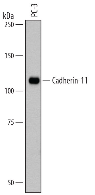
- Main image

- Experimental details
- Detection of Human Cadherin-11 by Western Blot. Western blot shows lysates of PC-3 human prostate cancer cell line. PVDF membrane was probed with 1 µg/mL of Mouse Anti-Human Cadherin-11 Monoclonal Antibody (Catalog # MAB1790) followed by HRP-conjugated Anti-Mouse IgG Secondary Antibody (Catalog # HAF007). A specific band was detected for Cadherin-11 at approximately 110 kDa (as indicated). This experiment was conducted under reducing conditions and using Immunoblot Buffer Group 1.
- Submitted by
- R&D Systems (provider)
- Main image
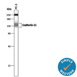
- Experimental details
- Detection of Human Cadherin-11 by Simple WesternTM. Simple Western lane view shows lysates of PC-3 human prostate cancer cell line, loaded at 0.2 mg/mL. Specific bands were detected for Cadherin-11 at approximately 114 & 137 kDa (as indicated) using 20 µg/mL of Mouse Anti-Human Cadherin-11 Monoclonal Antibody (Catalog # MAB1790). This experiment was conducted under reducing conditions and using the 12-230 kDa separation system. Non-specific interaction with the 230 kDa Simple Western standard may be seen with this antibody.

Supportive validation
- Submitted by
- R&D Systems (provider)
- Main image
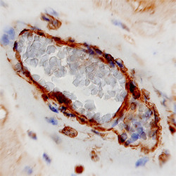
- Experimental details
- Cadherin-11 in Human Placenta. Cadherin-11 was detected in immersion fixed paraffin-embedded sections of human placenta using Mouse Anti-Human Cadherin-11 Monoclonal Antibody (Catalog # MAB1790) at 8 µg/mL overnight at 4 °C. Tissue was stained using the Anti-Mouse HRP-DAB Cell & Tissue Staining Kit (brown; Catalog # CTS002) and counterstained with hematoxylin (blue). View our protocol for Chromogenic IHC Staining of Paraffin-embedded Tissue Sections.
