Explore
 Validate
 Learn
LearnPA5-30220
antibody from Invitrogen Antibodies
Targeting: SRSF1
ASF, MGC5228, SF2, SF2p33, SFRS1, SRp30a
 Western blot
 Immunocytochemistry
Antibody data
- Antibody Data
- Antigen structure
- References [3]
- Comments [0]
- Validations
- Immunocytochemistry [1]
- Immunohistochemistry [4]
- Other assay [1]
Submit
Validation data
Reference
Comment
Report error
- Product number
- PA5-30220 - Provider product page

- Provider
- Invitrogen Antibodies
- Product name
- SRSF1 Polyclonal Antibody
- Antibody type
- Polyclonal
- Antigen
- Recombinant full-length protein
- Description
- Recommended positive controls: rat brain, HeLa, HepG2, Mouse brain. Predicted reactivity: Mouse (100%), Rat (100%), Pig (100%), Chicken (98%), Rhesus Monkey (100%), Chimpanzee (100%), Bovine (100%). Store product as a concentrated solution. Centrifuge briefly prior to opening the vial.
- Reactivity
- Human, Mouse, Rat
- Host
- Rabbit
- Isotype
- IgG
- Vial size
- 100 μL
- Concentration
- 1 mg/mL
- Storage
- Store at 4°C short term. For long term storage, store at -20°C, avoiding freeze/thaw cycles.
Submitted references An IGF-1R-mTORC1-SRPK2 signaling Axis contributes to FASN regulation in breast cancer.
Elucidating the Antiviral Mechanism of Different MARCH Factors.
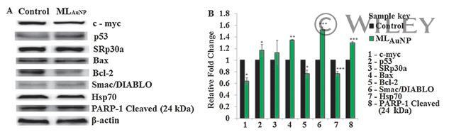
Moringa oleifera Gold Nanoparticles Modulate Oncogenes, Tumor Suppressor Genes, and Caspase-9 Splice Variants in A549 Cells.
McClellan B, Gries P, Harlow B, Tiziani S, Jolly C, deGraffenried L
BMC cancer 2022 Sep 12;22(1):976
BMC cancer 2022 Sep 12;22(1):976
Elucidating the Antiviral Mechanism of Different MARCH Factors.
Umthong S, Lynch B, Timilsina U, Waxman B, Ivey EB, Stavrou S
mBio 2021 Mar 2;12(2)
mBio 2021 Mar 2;12(2)
Moringa oleifera Gold Nanoparticles Modulate Oncogenes, Tumor Suppressor Genes, and Caspase-9 Splice Variants in A549 Cells.
Tiloke C, Phulukdaree A, Anand K, Gengan RM, Chuturgoon AA
Journal of cellular biochemistry 2016 Oct;117(10):2302-14
Journal of cellular biochemistry 2016 Oct;117(10):2302-14
No comments: Submit comment
Supportive validation
- Submitted by
- Invitrogen Antibodies (provider)
- Main image
- Experimental details
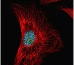
- Immunofluorescent analysis of SF2 in paraformaldehyde-fixed HeLa cells using a SF2 polyclonal antibody (Product # PA5-30220) (Green) at a 1:500 dilution. Alpha-tubulin filaments were labeled with Product # PA5-29281 (Red) at a 1:2000.

Supportive validation
- Submitted by
- Invitrogen Antibodies (provider)
- Main image
- Experimental details
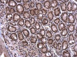
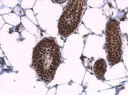
- SRSF1 Polyclonal Antibody detects SF2 protein at nucleus on mouse duodenum by immunohistochemical analysis. Sample: Paraffin-embedded mouse duodenum. SRSF1 Polyclonal Antibody (Product # PA5-30220) dilution: 1:500. Antigen Retrieval: EDTA based buffer, pH 8.0, 15 min.
- Submitted by
- Invitrogen Antibodies (provider)
- Main image
- Experimental details
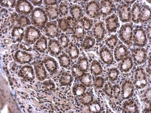
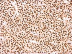
- SRSF1 Polyclonal Antibody detects SF2 protein at nucleus on mouse skin by immunohistochemical analysis. Sample: Paraffin-embedded mouse skin. SRSF1 Polyclonal Antibody (Product # PA5-30220) dilution: 1:500. Antigen Retrieval: EDTA based buffer, pH 8.0, 15 min.

- Submitted by
- Invitrogen Antibodies (provider)
- Main image
- Experimental details
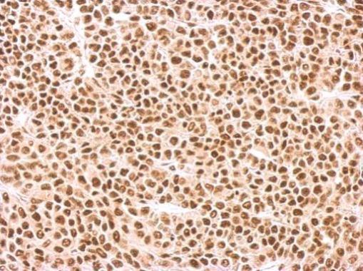
- SRSF1 Polyclonal Antibody detects SFRS1 protein at nucleus on BT483 xenograft by immunohistochemical analysis. Sample: Paraffin-embedded BT483 xenograft. SRSF1 Polyclonal Antibody (Product # PA5-30220) dilution: 1:500. Antigen Retrieval: EDTA based buffer, pH 8.0, 15 min.
- Submitted by
- Invitrogen Antibodies (provider)
- Main image
- Experimental details
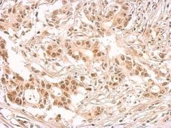
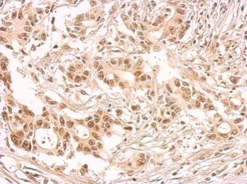
- SRSF1 Polyclonal Antibody detects SFRS1 protein at nucleus on human colon carcinoma by immunohistochemical analysis. Sample: Paraffin-embedded colon carcinoma. SRSF1 Polyclonal Antibody (Product # PA5-30220) dilution: 1:500. Antigen Retrieval: EDTA based buffer, pH 8.0, 15 min.
Supportive validation
- Submitted by
- Invitrogen Antibodies (provider)
- Main image
- Experimental details
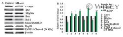
- The effect of ML AuNP on protein levels in A549 cells. Western blot analysis of protein levels (A) and the relative fold change (B) in A549 lung cancer cells after exposure to ML AuNP (*** P < 0.0001, ** P < 0.001, and * P < 0.05). Apoptotic proteins were significantly increased with a simultaneous decrease in anti-apoptotic proteins. Protein bands were normalized against beta-actin.