Explore
 Validate
 Learn
Learn Western blot
Antibody data
- Antibody Data
- Antigen structure
- References [8]
- Comments [0]
- Validations
- Western blot [4]
- Immunocytochemistry [5]
- Flow cytometry [1]
Submit
Validation data
Reference
Comment
Report error
- Product number
- 700184 - Provider product page

- Provider
- Invitrogen Antibodies
- Product name
- RAB11A Recombinant Rabbit Monoclonal Antibody (3H18L5)
- Antibody type
- Monoclonal
- Antigen
- Synthetic peptide
- Description
- This antibody is predicted to react with bovine, chicken, equine, mouse, primate, rat, Xenopus and zebrafish based on sequence homology.
- Antibody clone number
- 3H18L5
- Concentration
- 0.5 mg/mL
Submitted references TECPR1 promotes aggrephagy by direct recruitment of LC3C autophagosomes to lysosomes.
Human DEF6 deficiency underlies an immunodeficiency syndrome with systemic autoimmunity and aberrant CTLA-4 homeostasis.
Endosome-Mediated Epithelial Remodeling Downstream of Hedgehog-Gli Is Required for Tracheoesophageal Separation.
The extended cytoplasmic tail of the human B4GALNT2 is critical for its Golgi targeting and post-Golgi sorting.
Axoneme polyglutamylation regulated by Joubert syndrome protein ARL13B controls ciliary targeting of signaling molecules.
Ablation of SNX6 leads to defects in synaptic function of CA1 pyramidal neurons and spatial memory.
CCC- and WASH-mediated endosomal sorting of LDLR is required for normal clearance of circulating LDL.
Exogenous α-synuclein decreases raft partitioning of Cav2.2 channels inducing dopamine release.
Wetzel L, Blanchard S, Rama S, Beier V, Kaufmann A, Wollert T
Nature communications 2020 Jun 12;11(1):2993
Nature communications 2020 Jun 12;11(1):2993
Human DEF6 deficiency underlies an immunodeficiency syndrome with systemic autoimmunity and aberrant CTLA-4 homeostasis.
Serwas NK, Hoeger B, Ardy RC, Stulz SV, Sui Z, Memaran N, Meeths M, Krolo A, Yüce Petronczki Ö, Pfajfer L, Hou TZ, Halliday N, Santos-Valente E, Kalinichenko A, Kennedy A, Mace EM, Mukherjee M, Tesi B, Schrempf A, Pickl WF, Loizou JI, Kain R, Bidmon-Fliegenschnee B, Schickel JN, Glauzy S, Huemer J, Garncarz W, Salzer E, Pierides I, Bilic I, Thiel J, Priftakis P, Banerjee PP, Förster-Waldl E, Medgyesi D, Huber WD, Orange JS, Meffre E, Sansom DM, Bryceson YT, Altman A, Boztug K
Nature communications 2019 Jul 15;10(1):3106
Nature communications 2019 Jul 15;10(1):3106
Endosome-Mediated Epithelial Remodeling Downstream of Hedgehog-Gli Is Required for Tracheoesophageal Separation.
Nasr T, Mancini P, Rankin SA, Edwards NA, Agricola ZN, Kenny AP, Kinney JL, Daniels K, Vardanyan J, Han L, Trisno SL, Cha SW, Wells JM, Kofron MJ, Zorn AM
Developmental cell 2019 Dec 16;51(6):665-674.e6
Developmental cell 2019 Dec 16;51(6):665-674.e6
The extended cytoplasmic tail of the human B4GALNT2 is critical for its Golgi targeting and post-Golgi sorting.
Groux-Degroote S, Schulz C, Cogez V, Noël M, Portier L, Vicogne D, Solorzano C, Dall'Olio F, Steenackers A, Mortuaire M, Gonzalez-Pisfil M, Henry M, Foulquier F, Héliot L, Harduin-Lepers A
The FEBS journal 2018 Sep;285(18):3442-3463
The FEBS journal 2018 Sep;285(18):3442-3463
Axoneme polyglutamylation regulated by Joubert syndrome protein ARL13B controls ciliary targeting of signaling molecules.
He K, Ma X, Xu T, Li Y, Hodge A, Zhang Q, Torline J, Huang Y, Zhao J, Ling K, Hu J
Nature communications 2018 Aug 17;9(1):3310
Nature communications 2018 Aug 17;9(1):3310
Ablation of SNX6 leads to defects in synaptic function of CA1 pyramidal neurons and spatial memory.
Niu Y, Dai Z, Liu W, Zhang C, Yang Y, Guo Z, Li X, Xu C, Huang X, Wang Y, Shi YS, Liu JJ
eLife 2017 Jan 30;6
eLife 2017 Jan 30;6
CCC- and WASH-mediated endosomal sorting of LDLR is required for normal clearance of circulating LDL.
Bartuzi P, Billadeau DD, Favier R, Rong S, Dekker D, Fedoseienko A, Fieten H, Wijers M, Levels JH, Huijkman N, Kloosterhuis N, van der Molen H, Brufau G, Groen AK, Elliott AM, Kuivenhoven JA, Plecko B, Grangl G, McGaughran J, Horton JD, Burstein E, Hofker MH, van de Sluis B
Nature communications 2016 Mar 11;7:10961
Nature communications 2016 Mar 11;7:10961
Exogenous α-synuclein decreases raft partitioning of Cav2.2 channels inducing dopamine release.
Ronzitti G, Bucci G, Emanuele M, Leo D, Sotnikova TD, Mus LV, Soubrane CH, Dallas ML, Thalhammer A, Cingolani LA, Mochida S, Gainetdinov RR, Stephens GJ, Chieregatti E
The Journal of neuroscience : the official journal of the Society for Neuroscience 2014 Aug 6;34(32):10603-15
The Journal of neuroscience : the official journal of the Society for Neuroscience 2014 Aug 6;34(32):10603-15
No comments: Submit comment
Supportive validation
- Submitted by
- Invitrogen Antibodies (provider)
- Main image
- Experimental details
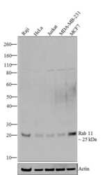
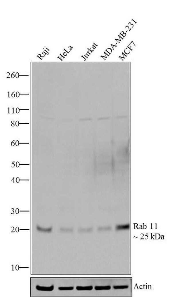
- Western blot analysis of Rab11 was performed by loading 20 µg of Raji (lane1), HeLa (lane2), Jurkat (lane3), MDA-MB-231 (lane4) and MCF7 (lane5) cell lysates using Novex®NuPAGE® 4-12% Bis-Tris gel (Product # NP0321BOX), XCell SureLock Electrophoresis System (Product # EI0002), Novex® Sharp Pre-Stained Protein Standard (Product # LC5800), and iBlot® Dry Blotting System (Product # IB21001). Proteins were transferred to a nitrocellulose membrane and blocked with 5 % skim milk for 1 hour at room temperature. Rab11 was detected at ~ 25 kDa using Rab11 Recombinant Rabbit Monoclonal Antibody (Product # 700184) at 1 µg-3 µg/mL in 2.5 % skim milk at 4°C overnight on a rocking platform. Goat anti-Rabbit IgG-HRP Secondary Antibody (Product # G-21234) at 1:5000 dilution was used and chemiluminescent detection was performed using Pierce™ ECL Western blotting Substrate (Product # 32106).
- Submitted by
- Invitrogen Antibodies (provider)
- Main image

- Experimental details
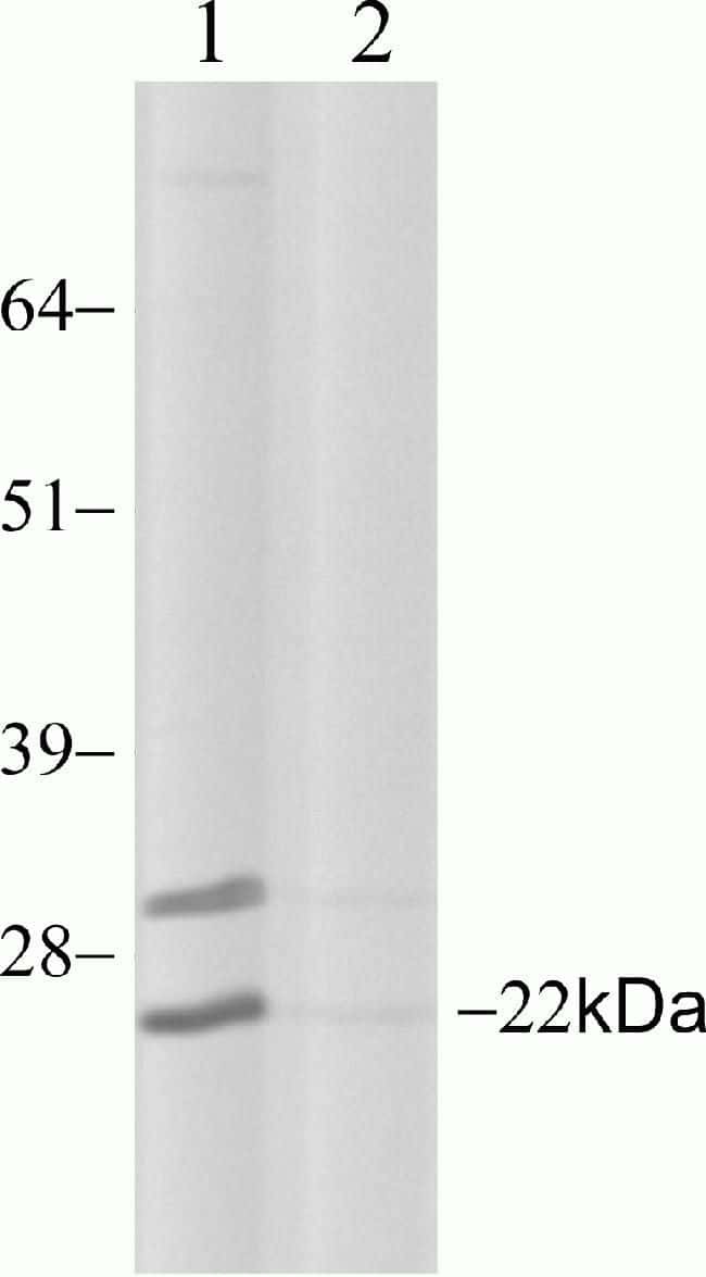
- Western blot analysis of Rab11 in HeLa cell lysate using a Rab11 recombinant rabbit monoclonal antibody (Product # 700184) at a dilution of 6.5 µg/mL.
- Submitted by
- Invitrogen Antibodies (provider)
- Main image
- Experimental details
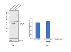
- Knockdown of Ras-related protein Rab-11A was achieved by transfecting HeLa with Ras-related protein Rab-11A specific siRNAs (Silencer® select Product # S16703, S16704). Western blot analysis (Fig. a) was performed using Membrane enriched extracts from the Ras-related protein Rab-11A knockdown cells (lane 3), non-targeting scrambled siRNA transfected cells (lane 2) and untransfected cells (lane 1). The blot was probed with RAB11A Recombinant Rabbit Monoclonal Antibody (3H18L5) (Product # 700184, 1 µg/mL) and Goat anti-Rabbit IgG (H+L) Superclonal™ Recombinant Secondary Antibody, HRP (Product # A27036, 1:20000 dilution). Densitometric analysis of this western blot is shown in histogram (Fig. b). Decrease in signal upon siRNA mediated knock down confirms that antibody is specific to Ras-related protein Rab-11A.

- Submitted by
- Invitrogen Antibodies (provider)
- Main image
- Experimental details
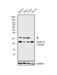
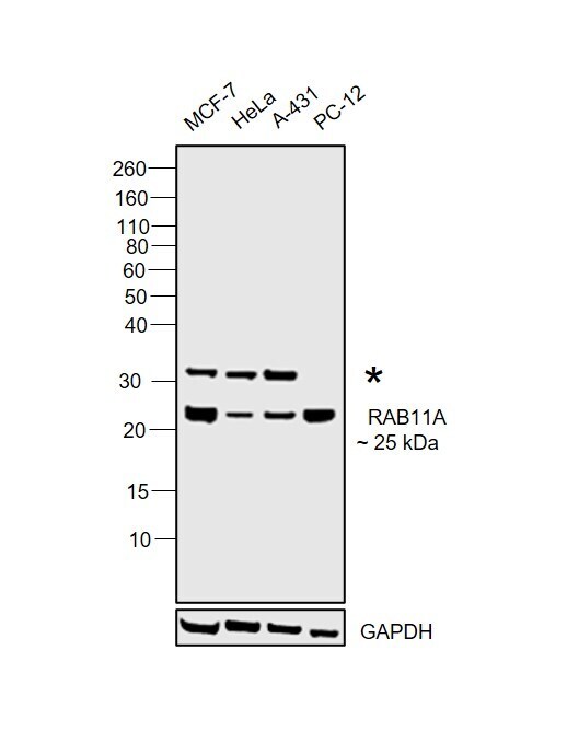
- Western blot was performed using Anti-RAB11A Recombinant Rabbit Monoclonal Antibody (3H18L5) (Product # 700184) and a 25 kDa band corresponding to Ras-related protein Rab-11A was observed across cell lines tested. An uncharacterised band (*) above 30 kDa was also observed across cell lines tested. Membrane enriched extracts (30 µg lysate) of MCF7 (Lane 1), HeLa (Lane 2), A-431 (Lane 3), and PC-12 (Lane 4) were electrophoresed using NuPAGE™ 12% Bis-Tris Protein Gel (Product # NP0342BOX). Resolved proteins were then transferred onto a nitrocellulose membrane (Product # IB23001) by iBlot® 2 Dry Blotting System (Product # IB21001). The blot was probed with the primary antibody (1 µg/mL) and detected by chemiluminescence with Goat anti-Rabbit IgG (H+L) Superclonal™ Recombinant Secondary Antibody, HRP (Product # A27036,1:20000 dilution) using the iBright FL 1000 (Product # A32752). Chemiluminescent detection was performed using SuperSignal™ West Pico PLUS Chemiluminescent Substrate (Product # 34580).
Supportive validation
- Submitted by
- Invitrogen Antibodies (provider)
- Main image
- Experimental details
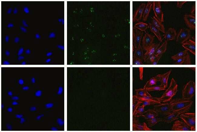
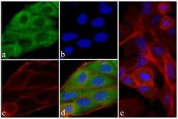
- Immunofluorescent analysis of Rab11 in HeLa cells using a Rab11 recombinant rabbit monoclonal antibody (Product # 700184) at a dilution of 2.5 µg/mL in the absence of peptide (top) and presence of immunogenic peptide (bottom), followed by detection using an Alexa Fluor 488-conjugated goat anti-rabbit secondary antibody at a dilution of 1:1000. Actin was stained with Alexa Fluor 568 Phalloidin (Product # A12380). Hoechst only (blue, left), AF488 signal only (green, middle) and composite image with Phalloidin (right).
- Submitted by
- Invitrogen Antibodies (provider)
- Main image

- Experimental details
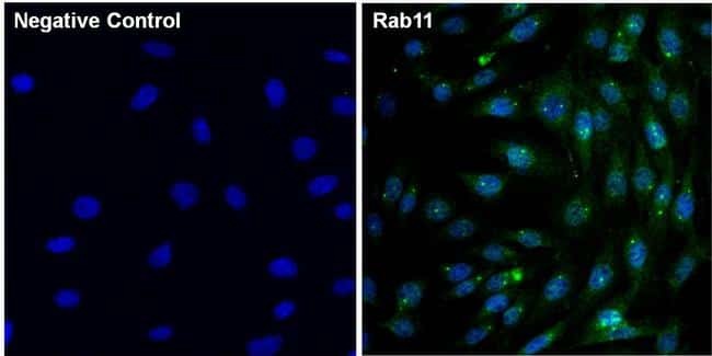
- Immunofluorescent analysis of Rab11 (green) in 3T3 cells. The cells were fixed with 4% paraformaldehyde for 15 minutes and blocked with 3% Blocker BSA (Product # 37525) in PBS for 30 minutes at room temperature. Cells were stained with or without Rab11 rabbit monoclonal antibody (Product # 700184), at a concentration of 5 µg/mL for 1 hour at room temperature, and then incubated with a Goat anti-Rabbit (H+L) Superclonal Secondary Antibody, Alexa Fluor® 488 conjugate (Product # A27034) at a dilution of 1:1000 for 1 hour at room temperature (both panels, green). Nuclei (both panels, blue) were stained with Hoechst 33342 dye (Product # 62249). Images were taken on a Thermo Scientific ToxInsight at 20X magnification.
- Submitted by
- Invitrogen Antibodies (provider)
- Main image
- Experimental details
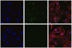
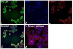
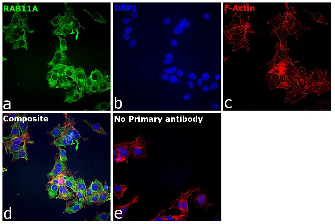
- Immunofluorescence analysis of Ras-related protein Rab-11A was performed using 70% confluent log phase A-431 cells. The cells were fixed with 4% paraformaldehyde for 10 minutes, permeabilized with 0.01% Triton™ X-100 for 15 minutes, and blocked with 2% BSA for 45 minutes at room temperature. The cells were labeled with RAB11A Recombinant Rabbit Monoclonal Antibody (3H18L5) (Product # 700184) at 1:100 in 0.1% BSA, incubated at 4 degree celsius overnight and then labeled with Donkey anti-Rabbit IgG (H+L) Highly Cross-Adsorbed Secondary Antibody, Alexa Fluor Plus 488 (Product # A32790), (1:2000), for 45 minutes at room temperature (Panel a: Green). Nuclei (Panel b:Blue) were stained with Hoechst 33342 (Product # H1399). F-actin (Panel c: Red) was stained with Rhodamine Phalloidin (Product # R415, 1:300). Panel d represents the merged image showing cytoplasmic localization. Panel e represents control cells with no primary antibody to assess background. The images were captured at 40X magnification in CellInsight CX7 LZR High-Content Screening (HCS) Platform (Product # CX7A1110LZR) and externally deconvoluted (D.Sage et al. / Methods 115 (2017) 28-41).

- Submitted by
- Invitrogen Antibodies (provider)
- Main image
- Experimental details
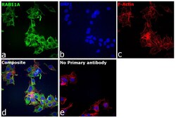
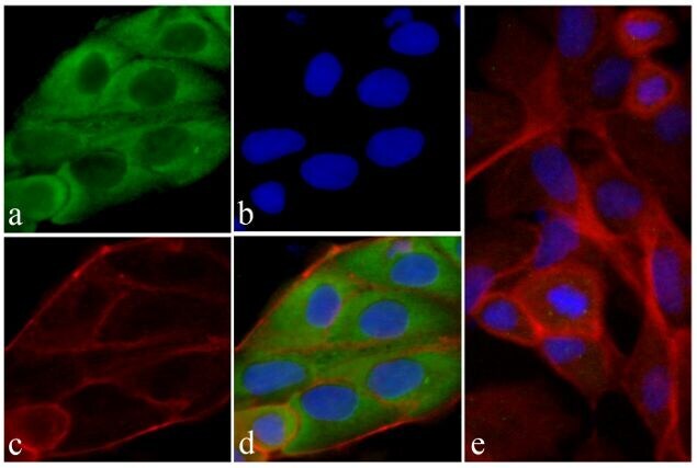
- Immunofluorescence analysis of Ras-related protein Rab-11A was performed using 70% confluent log phase Hep G2 cells. The cells were fixed with 4% paraformaldehyde for 10 minutes, permeabilized with 0.01% Triton™ X-100 for 15 minutes, and blocked with 2% BSA for 45 minutes at room temperature. The cells were labeled with RAB11A Polyclonal Antibody (Product # 700184) at 1:100 in 0.1% BSA, incubated at 4 degree celsius overnight and then labeled with Donkey anti-Rabbit IgG (H+L) Highly Cross-Adsorbed Secondary Antibody, Alexa Fluor Plus 488 (Product # A32790), (1:2,000), for 45 minutes at room temperature (Panel a: Green). Nuclei (Panel b:Blue) were stained with Hoechst 33342 (Product # H1399). F-actin (Panel c: Red) was stained with Rhodamine Phalloidin (Product # R415, 1:300). Panel d represents the merged image showing cytoplasmic localization. Panel e represents control cells with no primary antibody to assess background. The images were captured at 40X magnification in CellInsight CX7 LZR High-Content Screening (HCS) Platform (Product # CX7A1110LZR) and externally deconvoluted (D.Sage et al. / Methods 115 (2017) 28–41).
- Submitted by
- Invitrogen Antibodies (provider)
- Main image
- Experimental details
- Immunofluorescent analysis of Rab11 was done on 80% confluent log phase HeLa cells. The cells were fixed with 4% paraformaldehyde for 15 minutes; permeabilized with 0.25% Triton X-100 for 10 minutes followed by blocking with 5% BSA for 1 hour at room temperature. The cells were incubated with Rab11 Recombinant Rabbit Monoclonal Antibody (Product # 700184) at 2 µg-4 µg in 1% BSA and incubated for 3 hours at room temperature and then labeled with Alexa Fluor® 488 Goat anti-Rabbit IgG Secondary Antibody (Product # A-11008) at a dilution of 1:400 for 30 minutes at room temperature (Panel a: green). Nuclei (Panel b: blue) were stained with SlowFade® Gold Antifade Mountant with DAPI (Product # S36938). F-actin (Panel c: red) was stained with Alexa Fluor® 594 Phalloidin (Product # A12381). Panel d is a merged image showing cytoplasmic localization of Rab11. Panel e shows no primary antibody. The images were captured at 20X magnification.
Supportive validation
- Submitted by
- Invitrogen Antibodies (provider)
- Main image
- Experimental details
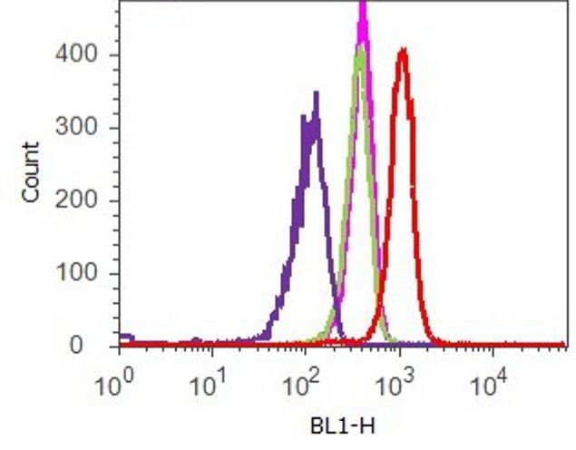
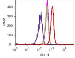
- Flow cytometry analysis of Rab11 was done on HeLa cells. Cells were fixed with 70% ethanol for 10 minutes, permeabilized with 0.25% Tritonª X-100 for 20 minutes, and blocked with 5% BSA for 1 hour at room temperature. Cells were labeled with ABfinityª Rab11 Recombinant Rabbit Monoclonal Antibody (700184, red histogram) or with rabbit isotype control (pink histogram) at 2 µg-4 µg/million cells in 2.5% BSA. After incubation at room temperature for 2-3 hours, the cells were labeled with Alexa Fluor¨ 488 Goat Anti-Rabbit Secondary Antibody (A11008) at a dilution of 1:400 for 30 minutes at room temperature. The representative 10,000 cells were acquired and analyzed for each sample using an Attune¨ Acoustic Focusing Cytometer. The purple histogram represents unstained control cells and the green histogram represents no-primary-antibody control.