 Explore
 Validate
 Learn
Learn Western blot
 Immunocytochemistry
Antibody data
- Antibody Data
- Antigen structure
- References [12]
- Comments [0]
- Validations
- Immunocytochemistry [1]
Submit
Validation data
Reference
Comment
Report error
- Product number
- HPA000866 - Provider product page

- Provider
- Atlas Antibodies
- Proper citation
- Atlas Antibodies Cat#HPA000866, RRID:AB_2276678
- Product name
- Anti-SYNJ2BP
- Antibody type
- Polyclonal
- Description
- Polyclonal Antibody against Human SYNJ2BP, Gene description: synaptojanin 2 binding protein, Alternative Gene Names: Arip2, Validated applications: ICC, IHC, WB, Uniprot ID: P57105, Storage: Store at +4°C for short term storage. Long time storage is recommended at -20°C.
- Reactivity
- Human, Mouse
- Host
- Rabbit
- Conjugate
- Unconjugated
- Isotype
- IgG
- Vial size
- 100 µl
- Concentration
- 0.1 mg/ml
- Storage
- Store at +4°C for short term storage. Long time storage is recommended at -20°C.
- Handling
- The antibody solution should be gently mixed before use.
Submitted references Protocol for measuring interorganelle contact sites in primary cells using a modified proximity ligation assay
Contact sites between endoplasmic reticulum sheets and mitochondria regulate mitochondrial DNA replication and segregation
Dynamic mapping of proteome trafficking within and between living cells by TransitID
Altered SYNJ2BP-mediated mitochondrial-ER contacts in motor neuron disease
Expression of Synj2bp in mouse liver regulates the extent of wrappER-mitochondria contact to maintain hepatic lipid homeostasis
Benchmarking a highly selective USP30 inhibitor for enhancement of mitophagy and pexophagy
Spatiotemporally-resolved mapping of RNA binding proteins via functional proximity labeling reveals a mitochondrial mRNA anchor promoting stress recovery
An engineered transcriptional reporter of protein localization identifies regulators of mitochondrial and ER membrane protein trafficking in high-throughput CRISPRi screens
USP30 sets a trigger threshold for PINK1–PARKIN amplification of mitochondrial ubiquitylation
The mitochondrial outer membrane protein SYNJ2BP interacts with the cell adhesion molecule TMIGD1 and can recruit it to mitochondria
Proteomic mapping of cytosol-facing outer mitochondrial and ER membranes in living human cells by proximity biotinylation
Tissue profiling of the mammalian central nervous system using human antibody-based proteomics.
Ilamathi H, Benhammouda S, Chatel-Chaix L, Germain M
STAR Protocols 2024;5(1):102915
STAR Protocols 2024;5(1):102915
Contact sites between endoplasmic reticulum sheets and mitochondria regulate mitochondrial DNA replication and segregation
Ilamathi H, Benhammouda S, Lounas A, Al-Naemi K, Desrochers-Goyette J, Lines M, Richard F, Vogel J, Germain M
iScience 2023;26(7):107180
iScience 2023;26(7):107180
Dynamic mapping of proteome trafficking within and between living cells by TransitID
Qin W, Cheah J, Xu C, Messing J, Freibaum B, Boeynaems S, Taylor J, Udeshi N, Carr S, Ting A
Cell 2023;186(15):3307-3324.e30
Cell 2023;186(15):3307-3324.e30
Altered SYNJ2BP-mediated mitochondrial-ER contacts in motor neuron disease
Pourshafie N, Masati E, Lopez A, Bunker E, Snyder A, Edwards N, Winkelsas A, Fischbeck K, Grunseich C
Neurobiology of Disease 2022;172
Neurobiology of Disease 2022;172
Expression of Synj2bp in mouse liver regulates the extent of wrappER-mitochondria contact to maintain hepatic lipid homeostasis
Ilacqua N, Anastasia I, Aloshyn D, Ghandehari-Alavijeh R, Peluso E, Brearley-Sholto M, Pellegrini L, Raimondi A, de Aguiar Vallim T, Pellegrini L
Biology Direct 2022;17(1)
Biology Direct 2022;17(1)
Benchmarking a highly selective USP30 inhibitor for enhancement of mitophagy and pexophagy
Rusilowicz-Jones E, Barone F, Lopes F, Stephen E, Mortiboys H, Urbé S, Clague M
Life Science Alliance 2021;5(2):e202101287
Life Science Alliance 2021;5(2):e202101287
Spatiotemporally-resolved mapping of RNA binding proteins via functional proximity labeling reveals a mitochondrial mRNA anchor promoting stress recovery
Qin W, Myers S, Carey D, Carr S, Ting A
Nature Communications 2021;12(1)
Nature Communications 2021;12(1)
An engineered transcriptional reporter of protein localization identifies regulators of mitochondrial and ER membrane protein trafficking in high-throughput CRISPRi screens
Yao D, Coukos R, Sanchez M, Strand E, Olive M, Udeshi N, Weissman J, Carr S, Bassik M, Ting A
eLife 2021;10
eLife 2021;10
USP30 sets a trigger threshold for PINK1–PARKIN amplification of mitochondrial ubiquitylation
Rusilowicz-Jones E, Jardine J, Kallinos A, Pinto-Fernandez A, Guenther F, Giurrandino M, Barone F, McCarron K, Burke C, Murad A, Martinez A, Marcassa E, Gersch M, Buckmelter A, Kayser-Bricker K, Lamoliatte F, Gajbhiye A, Davis S, Scott H, Murphy E, England K, Mortiboys H, Komander D, Trost M, Kessler B, Ioannidis S, Ahlijanian M, Urbé S, Clague M
Life Science Alliance 2020;3(8):e202000768
Life Science Alliance 2020;3(8):e202000768
The mitochondrial outer membrane protein SYNJ2BP interacts with the cell adhesion molecule TMIGD1 and can recruit it to mitochondria
Hartmann C, Schwietzer Y, Kummer D, Kirschnick N, Hoppe E, Thüring E, Glaesner-Ebnet M, Brinkmann F, Gerke V, Reuter S, Nakayama M, Ebnet K
BMC Molecular and Cell Biology 2020;21(1)
BMC Molecular and Cell Biology 2020;21(1)
Proteomic mapping of cytosol-facing outer mitochondrial and ER membranes in living human cells by proximity biotinylation
Hung V, Lam S, Udeshi N, Svinkina T, Guzman G, Mootha V, Carr S, Ting A
eLife 2017;6
eLife 2017;6
Tissue profiling of the mammalian central nervous system using human antibody-based proteomics.
Mulder J, Björling E, Jonasson K, Wernérus H, Hober S, Hökfelt T, Uhlén M
Molecular & cellular proteomics : MCP 2009 Jul;8(7):1612-22
Molecular & cellular proteomics : MCP 2009 Jul;8(7):1612-22
No comments: Submit comment
Supportive validation
- Submitted by
- Atlas Antibodies (provider)
- Main image
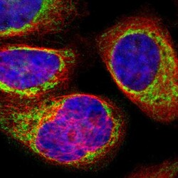
- Experimental details
- Immunofluorescent staining of human cell line A-431 shows localization to mitochondria.
- Sample type
- Human
