 Explore
 Validate
 Learn
Learn Western blot
 Immunocytochemistry
Antibody data
- Antibody Data
- Antigen structure
- References [2]
- Comments [0]
- Validations
- Western blot [10]
- Immunocytochemistry [2]
- Immunoprecipitation [1]
- Immunohistochemistry [5]
Submit
Validation data
Reference
Comment
Report error
- Product number
- GTX101310 - Provider product page

- Provider
- GeneTex
- Proper citation
- GeneTex Cat#GTX101310, RRID:AB_1949707
- Product name
- ATRX antibody [C3], C-term
- Antibody type
- Polyclonal
- Reactivity
- Human, Mouse, Rat
- Host
- Rabbit
Submitted references Paradoxical results obtained with Ki67-labeling and PHH3-mitosis index in glial tumors: a literature analysis.
Docetaxel-loaded solid lipid nanoparticles suppress breast cancer cells growth with reduced myelosuppression toxicity.
Elmaci İ, Altinoz MA, Bolukbasi FH, Yapicier O, Sav A
Clinical neuropathology 2017 Nov Dec;36(6):272-282
Clinical neuropathology 2017 Nov Dec;36(6):272-282
Docetaxel-loaded solid lipid nanoparticles suppress breast cancer cells growth with reduced myelosuppression toxicity.
Yuan Q, Han J, Cong W, Ge Y, Ma D, Dai Z, Li Y, Bi X
International journal of nanomedicine 2014;9:4829-46
International journal of nanomedicine 2014;9:4829-46
No comments: Submit comment
Enhanced validation
Supportive validation
- Submitted by
- GeneTex (provider)
- Enhanced method
- Genetic validation
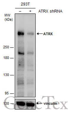
- Main image
- Experimental details
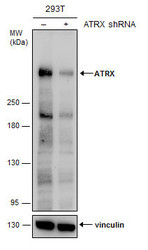
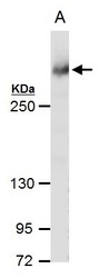
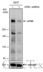
- Non-transfected (¡V) and transfected (+) 293T whole cell extracts (30 ?g) were separated by 5% SDS-PAGE, and the membrane was blotted with ATRX antibody [C3], C-term (GTX101310) diluted at 1:2000. The HRP-conjugated anti-rabbit IgG antibody (GTX213110-01) was used to detect the primary antibody.
Supportive validation
- Submitted by
- GeneTex (provider)
- Main image
- Experimental details
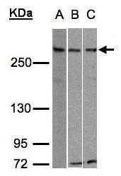
- Sample (30?g whole cell lysate)A:293TB:Hep G2 (GTX27900)C:Raji (GTX27908)5% SDS PAGEGTX101310 diluted at 1:500
- Validation comment
- WB

- Submitted by
- GeneTex (provider)
- Main image

- Experimental details
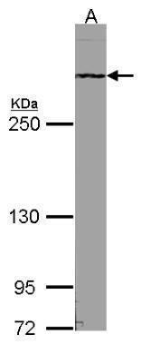
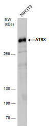
- Sample (30 ug of whole cell lysate) A:NIH-3T35% SDS PAGE GTX101310 diluted at 1:1000
- Validation comment
- WB
- Submitted by
- GeneTex (provider)
- Main image
- Experimental details
- Rad54 antibody [C3], C-term detects Rad54 protein by western blot analysis.A. 30 ?g NIH-3T3 whole cell extract5 % SDS-PAGERad54 antibody [C3], C-term (GTX101310) dilution: 1:1000
- Validation comment
- WB
- Submitted by
- GeneTex (provider)
- Main image
- Experimental details
- ATRX antibody detects ATRX protein by western blot analysis. Whole cell extracts (30 ?g) was separated by 5% SDS-PAGE, and the membrane was blotted with ATRX antibody (GTX101310) diluted by 1:1000. The HRP-conjugated anti-rabbit IgG antibody (GTX213110-01) was used to detect the primary antibody.

- Submitted by
- GeneTex (provider)
- Main image
- Experimental details
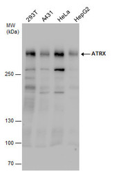
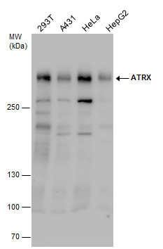
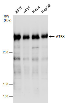
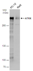
- ATRX antibody detects ATRX protein by western blot analysis. Various whole cell extracts (30 ?g) were separated by 5% SDS-PAGE, and the membrane was blotted with ATRX antibody (GTX101310) diluted by 1:1000.
- Validation comment
- WB
- Submitted by
- GeneTex (provider)
- Main image
- Experimental details
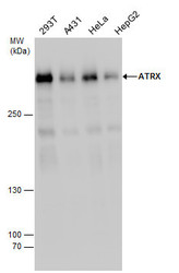
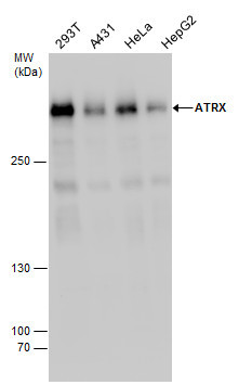
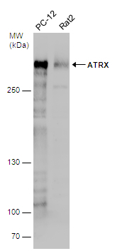
- ATRX antibody detects ATRX protein by western blot analysis. Various whole cell extracts (30 ?g) were separated by 5% SDS-PAGE, and the membrane was blotted with ATRX antibody (GTX101310) diluted at a dilution of 1:1000. The HRP-conjugated anti-rabbit IgG antibody (GTX213110-01) was used to detect the primary antibody.
- Submitted by
- GeneTex (provider)
- Main image
- Experimental details
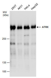
- Various whole cell extracts (30 ?g) were separated by 5% SDS-PAGE, and the membrane was blotted with ATRX antibody [C3], C-term (GTX101310) diluted at 1:1000. The HRP-conjugated anti-rabbit IgG antibody (GTX213110-01) was used to detect the primary antibody.
- Submitted by
- GeneTex (provider)
- Main image
- Experimental details
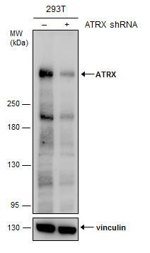
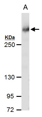
- Non-transfected (¡V) and transfected (+) 293T whole cell extracts (30 ?g) were separated by 5% SDS-PAGE, and the membrane was blotted with ATRX antibody [C3], C-term (GTX101310) diluted at 1:2000. The HRP-conjugated anti-rabbit IgG antibody (GTX213110-01) was used to detect the primary antibody.
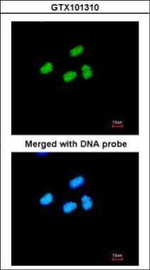
Supportive validation
- Submitted by
- GeneTex (provider)
- Main image
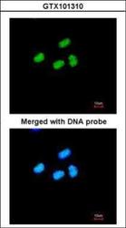
- Experimental details
- Immunofluorescence analysis of paraformaldehyde-fixed HeLa, using ATRX(GTX101310) antibody at 1:200 dilution.
- Submitted by
- GeneTex (provider)
- Main image
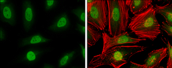
- Experimental details
- ATRX antibody [C3], C-term detects ATRX protein at nucleus by immunofluorescent analysis.Sample: HeLa cells were fixed in 4% paraformaldehyde at RT for 15 min.Green: ATRX stained by ATRX antibody [C3], C-term (GTX101310) diluted at 1:500.Red: phalloidin, a cytoskeleton marker, diluted at 1:100.

Supportive validation
- Submitted by
- GeneTex (provider)
- Main image
- Experimental details
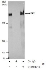
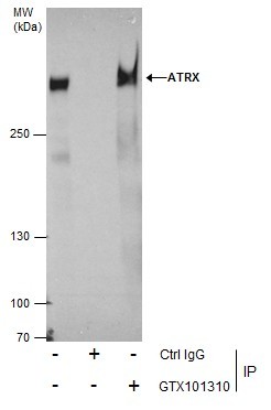
- Immunoprecipitation of ATRX protein from 293T whole cell extracts using 5 £gg of ATRX antibody [C3], C-term (GTX101310).Western blot analysis was performed using ATRX antibody [C3], C-term (GTX101310).EasyBlot anti-Rabbit IgG (GTX221666-01) was used as a secondary reagent.
Supportive validation
- Submitted by
- GeneTex (provider)
- Main image

- Experimental details
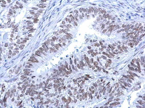
- Rad54 antibody [C3], C-term detects Rad54 protein at nucleus on human lung carcinoma by immunohistochemical analysis. Sample: Paraffin-embedded human lung carcinoma. Rad54 antibody [C3], C-term (GTX101310) dilution: 1:500.
- Submitted by
- GeneTex (provider)
- Main image

- Experimental details
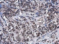
- Rad54 antibody [C3], C-term detects Rad54 protein at nucleus on human breast carcinoma by immunohistochemical analysis. Sample: Paraffin-embedded human breast carcinoma. Rad54 antibody [C3], C-term (GTX101310) dilution: 1:500.
- Submitted by
- GeneTex (provider)
- Main image
- Experimental details
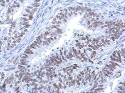
- Rad54 antibody [C3], C-term detects Rad54 protein at nucleus on human endometrial carcinoma by immunohistochemical analysis. Sample: Paraffin-embedded human endometrial carcinoma. Rad54 antibody [C3], C-term (GTX101310) dilution: 1:500.
- Submitted by
- GeneTex (provider)
- Main image
- Experimental details
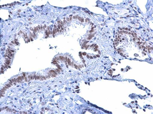
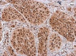
- ATRX antibody [C3], C-term detects ATRX protein at cytoplasm and nucleus by immunohistochemical analysis.Sample: Paraffin-embedded human breast carcinoma.ATRX stained by ATRX antibody [C3], C-term (GTX101310) diluted at 1:2000.

- Submitted by
- GeneTex (provider)
- Main image
- Experimental details
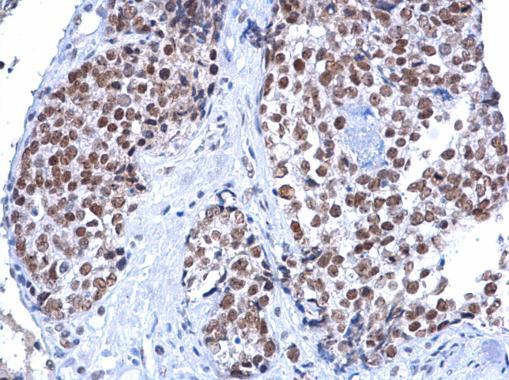
- ATRX antibody [C3], C-term detects ATRX protein at nucleus by immunohistochemical analysis.Sample: Paraffin-embedded human breast carcinoma.ATRX stained by ATRX antibody [C3], C-term (GTX101310) diluted at 1:2000.
