Explore
 Validate
 Learn
Learn Western blot
 Immunocytochemistry
Antibody data
- Antibody Data
- Antigen structure
- References [1]
- Comments [0]
- Validations
- Immunocytochemistry [2]
- Immunohistochemistry [4]
- Other assay [1]
Submit
Validation data
Reference
Comment
Report error
- Product number
- PA5-21348 - Provider product page

- Provider
- Invitrogen Antibodies
- Product name
- ATRX Polyclonal Antibody
- Antibody type
- Polyclonal
- Antigen
- Recombinant full-length protein
- Description
- Recommended positive controls: 293T, A431, HeLa, HepG2, NIH3T3, PC-12, Rat-2. Predicted reactivity: Mouse (97%), Chimpanzee (100%). Store product as a concentrated solution. Centrifuge briefly prior to opening the vial.
- Reactivity
- Human, Mouse, Rat
- Host
- Rabbit
- Isotype
- IgG
- Vial size
- 100 μL
- Concentration
- 0.27 mg/mL
- Storage
- Store at 4°C short term. For long term storage, store at -20°C, avoiding freeze/thaw cycles.
Submitted references ATRX proximal protein associations boast roles beyond histone deposition.
Scott WA, Dhanji EZ, Dyakov BJA, Dreseris ES, Asa JS, Grange LJ, Mirceta M, Pearson CE, Stewart GS, Gingras AC, Campos EI
PLoS genetics 2021 Nov;17(11):e1009909
PLoS genetics 2021 Nov;17(11):e1009909
No comments: Submit comment
Supportive validation
- Submitted by
- Invitrogen Antibodies (provider)
- Main image
- Experimental details
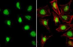
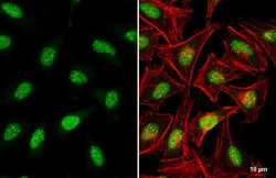
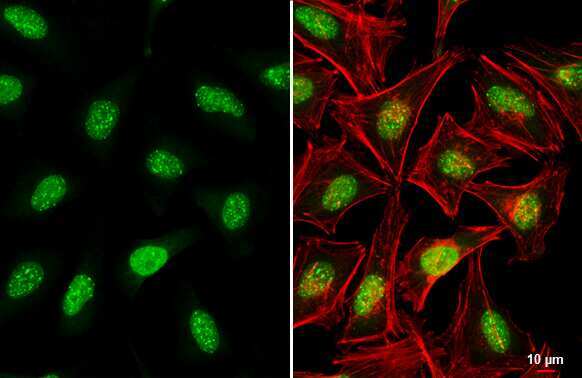
- Immunocytochemistry-Immunofluorescence analysis of ATRX was performed in HeLa cells fixed in 4% paraformaldehyde at RT for 15 min. Green: ATRX Polyclonal Antibody (Product # PA5-21348) diluted at 1:1500. Red: phalloidin, a cytoskeleton marker.
- Submitted by
- Invitrogen Antibodies (provider)
- Main image
- Experimental details
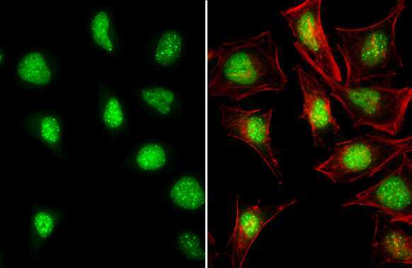
- ATRX Polyclonal Antibody detects ATRX protein at nucleus by immunofluorescent analysis. Sample: HeLa cells were fixed in 4% paraformaldehyde at RT for 15 min. Green: ATRX stained by ATRX Polyclonal Antibody (Product # PA5-21348) diluted at 1:1,000. Red: phalloidin, a cytoskeleton marker, diluted at 1:200. Scale bar= 10 µm.
Supportive validation
- Submitted by
- Invitrogen Antibodies (provider)
- Main image
- Experimental details
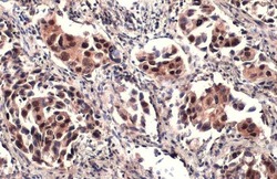
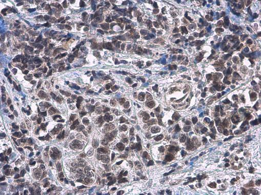
- ATRX Polyclonal Antibody detects ATRX protein at cytoplasm and nucleus by immunohistochemical analysis. Sample: Paraffin-embedded human lung cancer. ATRX stained by ATRX Polyclonal Antibody (Product # PA5-21348) diluted at 1:2,500. Antigen Retrieval: Citrate buffer, pH 6.0, 15 min.

- Submitted by
- Invitrogen Antibodies (provider)
- Main image
- Experimental details
- Rad54 antibody [C3], C-term detects Rad54 protein at nucleus on human endometrial carcinoma by immunohistochemical analysis. Sample: Paraffin-embedded human endometrial carcinoma. Rad54 antibody [C3], C-term (Product # PA5-21348) dilution: 1:500.

- Submitted by
- Invitrogen Antibodies (provider)
- Main image
- Experimental details
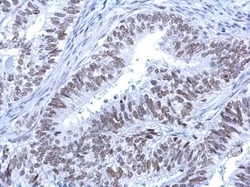
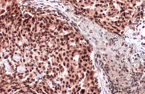
- Immunohistochemistry (Paraffin) analysis of ATRX was performed in paraffin-embedded human breast carcinoma tissue using ATRX Polyclonal Antibody (Product # PA5-21348) at a dilution of 1:2000.
- Submitted by
- Invitrogen Antibodies (provider)
- Main image

- Experimental details
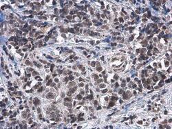
- Immunohistochemistry (Paraffin) analysis of ATRX was performed in paraffin-embedded human lung cancer tissue using ATRX Polyclonal Antibody (Product # PA5-21348) at a dilution of 1:500. Antigen Retrieval: Citrate buffer, pH 6.0, 15 min.
Supportive validation
- Submitted by
- Invitrogen Antibodies (provider)
- Main image

- Experimental details
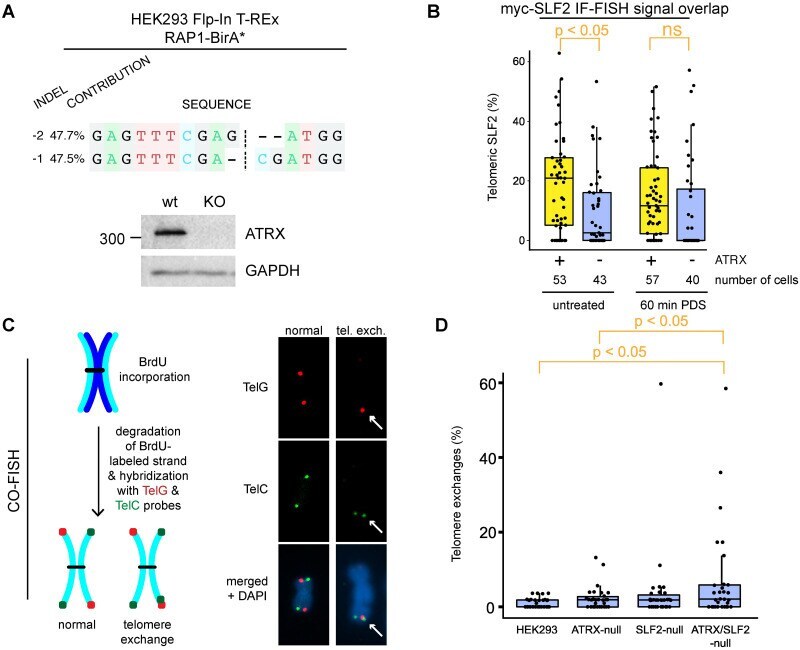
- 10.1371/journal.pgen.1009909.g004 Fig 4 Loss of ATRX and SLF2 enables telomere exchanges. A: DNA sequencing and western blotting confirming the disruption of ATRX expression in CRISPR-Cas9-edited HEK293 Flp-In T-REx cells inducibly expressing RAP1-BirA*. B: Relative signal overlap between immunolabeled SLF2 and telomere fluorescence in situ hybridization (IF-FISH), in ATRX-expressing or - HEK293 Flp-In cells. Cells were exposed to 10 muM pyridostatin (PDS, G4 stabilizer) for 60 min. At least 40 cells per condition were analyzed using a CellProfiler colocalization pipeline []. ATRX loss decreased SLF2 recruitment to telomeres in the absence of pyridostatin. C: Schematic of dual-colour chromosome-orientation fluorescence in situ hybridization (CO-FISH) [] using PNA probes to label C- and G-rich telomere strands (left). Examples of normal CO-FISH signals and of a chromosome end with a telomere exchange are shown (right). D: Relative amount of telomere exchanges in ATRX, SLF2, and ATRX/SLF2 KO HEK293 Flp-In cells. At least 30 mitotic spreads per condition were counted and the percent of telomeric exchanges plotted. p-values were obtained using a 2-sided Student's t-test with unequal variance.