 Explore
 Validate
 Learn
Learn Western blot
Antibody data
- Antibody Data
- Antigen structure
- References [1]
- Comments [0]
- Validations
- Western blot [2]
- ELISA [1]
- Immunocytochemistry [1]
Submit
Validation data
Reference
Comment
Report error
- Product number
- 701147 - Provider product page

- Provider
- Invitrogen Antibodies
- Product name
- PAX3 Recombinant Rabbit Monoclonal Antibody (16H22L10)
- Antibody type
- Monoclonal
- Antigen
- Recombinant full-length protein
- Description
- This antibody is predicted to react with mouse based on sequence homology.
- Antibody clone number
- 16H22L10
- Concentration
- 0.5 mg/mL
Submitted references BRN2 expression increases anoikis resistance in melanoma.
Pierce CJ, Simmons JL, Broit N, Karunarathne D, Ng MF, Boyle GM
Oncogenesis 2020 Jul 6;9(7):64
Oncogenesis 2020 Jul 6;9(7):64
No comments: Submit comment
Supportive validation
- Submitted by
- Invitrogen Antibodies (provider)
- Main image
- Experimental details
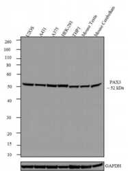
- Western blot analysis was performed on whole cell extracts (30 µg lysate) of U2OS (Lane 1), A431 (Lane 2), A375 (Lane 3), HEK-293 (Lane 4), THP1 (Lane 5), Mouse Testis (Lane 6) and Mouse Cerebellum (Lane 7). The blots were probed with Recombinant Rabbit Monoclonal Anti-PAX3 Antibody (Product # 701147, 1-2 µg/mL) and detected by chemiluminescence using Goat anti-Rabbit IgG (H+L) Recombinant Superclonal™ Secondary Antibody, HRP conjugate (Product # A27036, 0.4 µg/mL, 1:2500 dilution). A ~ 52 kDa band corresponding to PAX3 was observed across cell lines tested.Known quantity of protein samples were electrophoresed using Novex® NuPAGE® 4-12 % Bis-Tris gel (Product # NP0321BOX), XCell SureLock™ Electrophoresis System (Product # EI0002) and Novex® Sharp Pre-Stained Protein Standard (Product # LC5800). Resolved proteins were then transferred onto a nitrocellulose membrane with Pierce™ Power Blotter System (Product # 22834). The membrane was probed with the relevant primary and secondary Antibody following blocking with 5 % skimmed milk. Chemiluminescent detection was performed using Pierce™ ECL Western blotting Substrate (Product # 32106).

- Submitted by
- Invitrogen Antibodies (provider)
- Main image
- Experimental details
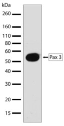
- Western blot analysis of PAX3 in A431 whole cell extracts using a PAX3 recombinant rabbit monoclonal antibody (Product # 701147) at a dilution of 2 µg/mL. Samples were detected using chemiluminescence (ECL). Results show a band at ~53kDa.

Supportive validation
- Submitted by
- Invitrogen Antibodies (provider)
- Main image

- Experimental details
- Indirect ELISA analysis of PAX3 in whole cell extracts of A431 coated onto the plate using a PAX3 recombinant rabbit monoclonal antibody (Product # 701147) at various dilutions. A non-linear regression analysis was performed (4 PL) and LOD and LOQ for the antibody were determined.

Supportive validation
- Submitted by
- Invitrogen Antibodies (provider)
- Main image

- Experimental details
- Immunofluorescent analysis of PAX3 in U2OS cells using a PAX3 recombinant rabbit monoclonal antibody (Product # 701147) followed by detection using an Alexa Fluor 488-conjugated goat anti-rabbit secondary antibody (green) (Image A). Nuclei were stained using DAPI (Image B) and actin stained with Alexa Fluor 594 phalloidin (red) (image C). Image D is a composite image showing nuclear localization of Pax 3.
