Explore
 Validate
 Learn
Learn Western blot
Antibody data
- Antibody Data
- Antigen structure
- References [3]
- Comments [0]
- Validations
- Western blot [2]
- Immunocytochemistry [2]
- Immunoprecipitation [1]
- Immunohistochemistry [2]
- Other assay [6]
Submit
Validation data
Reference
Comment
Report error
- Product number
- PA1-107 - Provider product page

- Provider
- Invitrogen Antibodies
- Product name
- PAX3 Polyclonal Antibody
- Antibody type
- Polyclonal
- Antigen
- Other
- Description
- PA1-107 has successfully been used in Western blot for detecting overexpressed and recombinant human Pax3 protein, but may not be suitable for detecting endogenous Pax3. By immunofluorescence, PA1-107 has been used to stain frozen embryonic mouse brain tissue. PA1-107 has been validated for IP using overexpressed Pax3 protein.
- Reactivity
- Human, Mouse
- Host
- Rabbit
- Isotype
- IgG
- Vial size
- 100 μg
- Concentration
- 1 mg/mL
- Storage
- -20°C
Submitted references [Screening of Mothers in a COVID-19 Low-Prevalence Region: Determination of SARS-CoV-2 Antibodies in 401 Mothers from Rostock by ELISA and Confirmation by Immunofluorescence].
Netrin1/DCC signaling promotes neuronal migration in the dorsal spinal cord.
NOVA regulates Dcc alternative splicing during neuronal migration and axon guidance in the spinal cord.
Reisinger EC, von Possel R, Warnke P, Geerdes-Fenge HF, Hemmer CJ, Pfefferle S, Löbermann M, Littmann M, Emmerich P
Deutsche medizinische Wochenschrift (1946) 2020 Aug;145(17):e96-e100
Deutsche medizinische Wochenschrift (1946) 2020 Aug;145(17):e96-e100
Netrin1/DCC signaling promotes neuronal migration in the dorsal spinal cord.
Junge HJ, Yung AR, Goodrich LV, Chen Z
Neural development 2016 Oct 26;11(1):19
Neural development 2016 Oct 26;11(1):19
NOVA regulates Dcc alternative splicing during neuronal migration and axon guidance in the spinal cord.
Leggere JC, Saito Y, Darnell RB, Tessier-Lavigne M, Junge HJ, Chen Z
eLife 2016 May 25;5
eLife 2016 May 25;5
No comments: Submit comment
Supportive validation
- Submitted by
- Invitrogen Antibodies (provider)
- Main image
- Experimental details
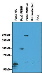
- Western blot analysis of Pax3 was performed by loading 40 µL of 500 µL RIPA cell extracts originating from 35mm dishes of HeLa cells transfected for 24 hours with the indicated Pax3-fusion constructs, untransfected HeLa cells, or Rh3 alveolar rhabdomyosarcoma cells per well onto an 8% Tris-Glycine gel. Proteins were transferred to a nitrocellulose membrane, and blocked with 2% BSA in PBST for 10 minutes at room temperature. Pax3 was detected at various molecular weights (dictated by the size of the gene fusion) using a Pax3 polyclonal antibody at a concentration of 1 µg/mL overnight at 4C, followed by an HRP-conjugated anti-rabbit IgG. Chemiluminescent detection was performed using ECL substrate. Data supplied courtesy of the Innovator's Program.

- Submitted by
- Invitrogen Antibodies (provider)
- Main image
- Experimental details
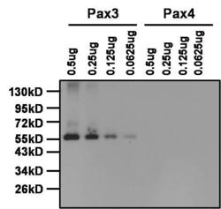
- Western blot analysis of Pax3 was performed by loading the indicated amounts of Pax3 and Pax4 (negative control) recombinant proteins, and 10 µL of PageRuler Prestained Protein Ladder (Product # 26616) per well onto a Novex® 4-20% Tris-Glycine polyacrylamide gel. Proteins were transferred to a PVDF membrane using the G2 Fast Blotter (Product # 62288), and blocked with 5% milk in TBST for at least 1 hour at room temperature. Pax3 was detected at ~53 kD using a Pax3 polyclonal antibody (Product # PA1-107) at a dilution of 1:1000 in blocking buffer overnight at 4C on a rocking platform, followed by an HRP-conjugated goat anti-rabbit IgG secondary antibody (Product # 31460) at a dilution of 1:40,000 for at least 30 minutes. Chemiluminescent detection was performed using SuperSignal West Pico (Product # 34080). NOTE: The presence of Pax4 recombinant protein was determined by probing a duplicate blot with a Pax4 polyclonal antibody (Product # PA1-108), and the corresponding image is on the webpage for Product # PA1-108.

Supportive validation
- Submitted by
- Invitrogen Antibodies (provider)
- Main image
- Experimental details
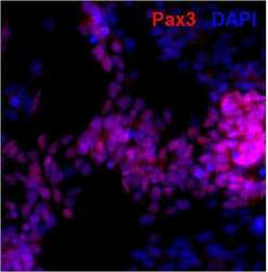
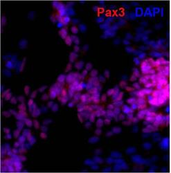
- Immunofluorescent analysis of Pax3 (red) in human neural stem cells derived from PD-3 iPSCs using Gibco® PSC Neural Induction Medium (Product # A1647801). The cells were fixed and permeabilized using Image-IT® Fixation/Permeabilization kit (Product # R37602), and blocked with blocking buffer included the kit for one hour at room temperature. Cells were stained with a Pax3 polyclonal antibody (Product # PA1-107) at a dilution of 1:200 in blocking buffer for 3 hours at room temperature, and then incubated with an AlexaFluor 594- conjugated donkey anti-rabbit IgG secondary antibody (Product # A-21207) at a dilution of 1:250 for 1 hour at room temperature. Nuclei (blue) were stained with DAPI (Product # D1306). Images were taken on an EVOS® FLoid® Cell Imaging Station at 10X magnification.

- Submitted by
- Invitrogen Antibodies (provider)
- Main image
- Experimental details
- Immunofluorescent analysis of Pax3 (red) in human neural stem cells derived from PD-3 iPSCs using Gibco® PSC Neural Induction Medium (Product # A1647801). The cells were fixed and permeabilized using Image-IT® Fixation/Permeabilization kit (Product # R37602), and blocked with blocking buffer included the kit for one hour at room temperature. Cells were stained with a Pax3 polyclonal antibody (Product # PA1-107) at a dilution of 1:200 in blocking buffer for 3 hours at room temperature, and then incubated with an AlexaFluor 594- conjugated donkey anti-rabbit IgG secondary antibody (Product # A-21207) at a dilution of 1:250 for 1 hour at room temperature. Nuclei (blue) were stained with DAPI (Product # D1306). Images were taken on an EVOS® FLoid® Cell Imaging Station at 10X magnification.

Supportive validation
- Submitted by
- Invitrogen Antibodies (provider)
- Main image
- Experimental details
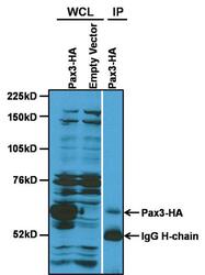
- Immunoprecipitation of Pax3 was performed on RIPA cell extracts originating from a 6cm dish of HeLa cells transfected for 24 hours with a Pax3-HA construct. Cells were lysed in 1 mL of RIPA buffer, and antigen-antibody complexes were formed by incubating 225 µL of HeLa Pax-HA cell extract with 2 µg of a Pax3 polyclonal antibody (Product # PA1-107) and Protein A sepharose for 4 hours at 4C. The samples were washed extensively with RIPA buffer, and eluted by boiling in SDS sample buffer. Samples were resolved on an 8% Tris-Glycine gel alongside whole cell lysate (WCL) from empty vector and Pax3-HA-transfected HeLa cells, transferred to a nitrocellulose membrane, and blocked with 2% BSA in PBST for 10 minutes at room temperature. The membrane was probed with an anti-HA polyclonal antibody overnight at 4C, washed in TBST, and probed with an HRP-conjugated anti-rabbit IgG secondary antibody. Chemiluminescent detection was performed using ECL substrate. Data supplied courtesy of the Innovator's Program.

Supportive validation
- Submitted by
- Invitrogen Antibodies (provider)
- Main image

- Experimental details
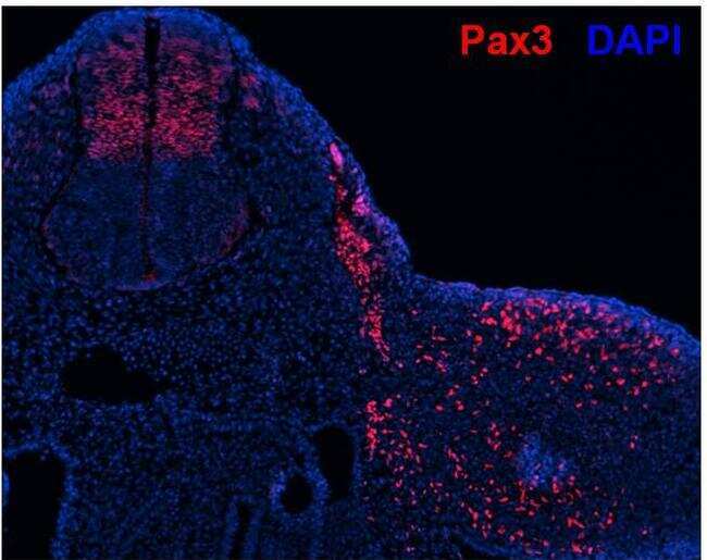
- Immunofluorescence was performed on mouse E10.5 embryo trunk at the forelimb level. Embryos were fixed with 4%PFA in 0.1M phosphate buffer, pH 7.2 at 4C for 30 minutes, processed for cryoprotection in 20% sucrose, and embedded in OCT compound for cryosectioning at 10 µm thickness. Following sectioning, tissues were washed in PBS with 0.1% Triton-X 100 (PBT), blocked in 1% goat serum in PBT for 30 minutes at room temperature, and then probed with a Pax3 polyclonal antibody (Product # PA1-107) at a concentration of 1 µg/mL in blocking buffer overnight at 4C. Tissues were washed extensively with PBT, and detection was performed using an Alexa Fluor 568-conjugated goat anti-rabbit IgG secondary antibody (red) at a dilution of 1:1000 in blocking buffer. Nuclei (blue) were stained with DAPI. Note: Staining was observed in the dorsal neural tube (symmetric two halves on the left side of the image), the dermomyotome/myotome (middle streak between symmetric halves), and limb muscle progenitors (scattered cells to the right side of the image). Data supplied courtesy of the Innovator's Program.
- Submitted by
- Invitrogen Antibodies (provider)
- Main image

- Experimental details
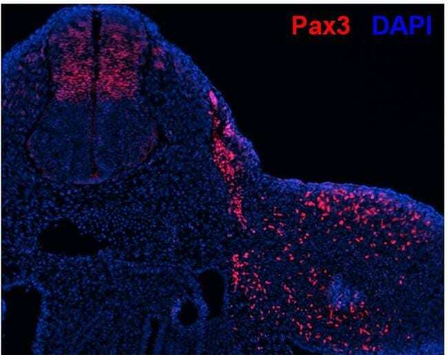
- Immunofluorescence was performed on mouse E10.5 embryo trunk at the forelimb level. Embryos were fixed with 4%PFA in 0.1M phosphate buffer, pH 7.2 at 4C for 30 minutes, processed for cryoprotection in 20% sucrose, and embedded in OCT compound for cryosectioning at 10 µm thickness. Following sectioning, tissues were washed in PBS with 0.1% Triton-X 100 (PBT), blocked in 1% goat serum in PBT for 30 minutes at room temperature, and then probed with a Pax3 polyclonal antibody (Product # PA1-107) at a concentration of 1 µg/mL in blocking buffer overnight at 4C. Tissues were washed extensively with PBT, and detection was performed using an Alexa Fluor 568-conjugated goat anti-rabbit IgG secondary antibody (red) at a dilution of 1:1000 in blocking buffer. Nuclei (blue) were stained with DAPI. Note: Staining was observed in the dorsal neural tube (symmetric two halves on the left side of the image), the dermomyotome/myotome (middle streak between symmetric halves), and limb muscle progenitors (scattered cells to the right side of the image). Data supplied courtesy of the Innovator's Program.
Supportive validation
- Submitted by
- Invitrogen Antibodies (provider)
- Main image

- Experimental details
- Immunoprecipitation of Pax3 was performed on RIPA cell extracts originating from a 6cm dish of HeLa cells transfected for 24 hours with a Pax3-HA construct. Cells were lysed in 1 mL of RIPA buffer, and antigen-antibody complexes were formed by incubating 225 µL of HeLa Pax-HA cell extract with 2 µg of a Pax3 polyclonal antibody (Product # PA1-107) and Protein A sepharose for 4 hours at 4C. The samples were washed extensively with RIPA buffer, and eluted by boiling in SDS sample buffer. Samples were resolved on an 8% Tris-Glycine gel alongside whole cell lysate (WCL) from empty vector and Pax3-HA-transfected HeLa cells, transferred to a nitrocellulose membrane, and blocked with 2% BSA in PBST for 10 minutes at room temperature. The membrane was probed with an anti-HA polyclonal antibody overnight at 4C, washed in TBST, and probed with an HRP-conjugated anti-rabbit IgG secondary antibody. Chemiluminescent detection was performed using ECL substrate. Data supplied courtesy of the Innovator's Program.

- Submitted by
- Invitrogen Antibodies (provider)
- Main image
- Experimental details
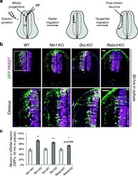
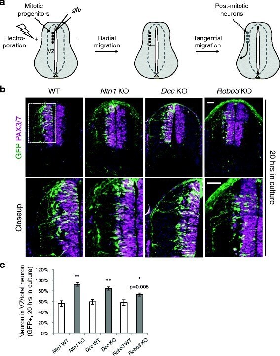
- Fig. 1 Neuroprogenitor migration in Ntn1 , Dcc , and Robo3 knockouts. a Schematic of the migration of the dorsal spinal cord progenitors and interneurons. VZ, ventricular zone. b Cross sections of the spinal cord electroporated with Actb-gfp ( betaactin-gfp) . The closeup images are of the boxed area. The embryos were cultured for 20 h. GFP+ neurons from all three KOs migrate out of the VZ (demarcated by PAX3/7 staining) later than WT neurons. c Quantification of the ratio between GFP+ neurons within the VZ and the total GFP+ neurons. A higher percentage of neurons is seen within the VZ in all three KOs. Data are represented as the mean +- SEM (Student's t -test; **, p < 0.001; *, p < 0.05). Scale bars, 50 mum
- Submitted by
- Invitrogen Antibodies (provider)
- Main image
- Experimental details
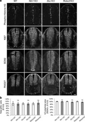
- Fig. 4 Neuroprogenitors are generated normally in Ntn1 , Dcc , and Robo3 knockouts. a Immunohistochemistry of phospho-Histone H3, a mitotic marker, Ki67, a cell proliferation marker, SOX2, a neuroprogenitor marker, and PAX3/7, a dorsal progenitor marker in E10.5 spinal cord. b Quantification of phenotypes in (a). Data are normalized to WT and are represented as the mean +- SEM (Student's t -test; ns, not significant). Scale bar, 50 mum

- Submitted by
- Invitrogen Antibodies (provider)
- Main image
- Experimental details
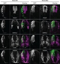
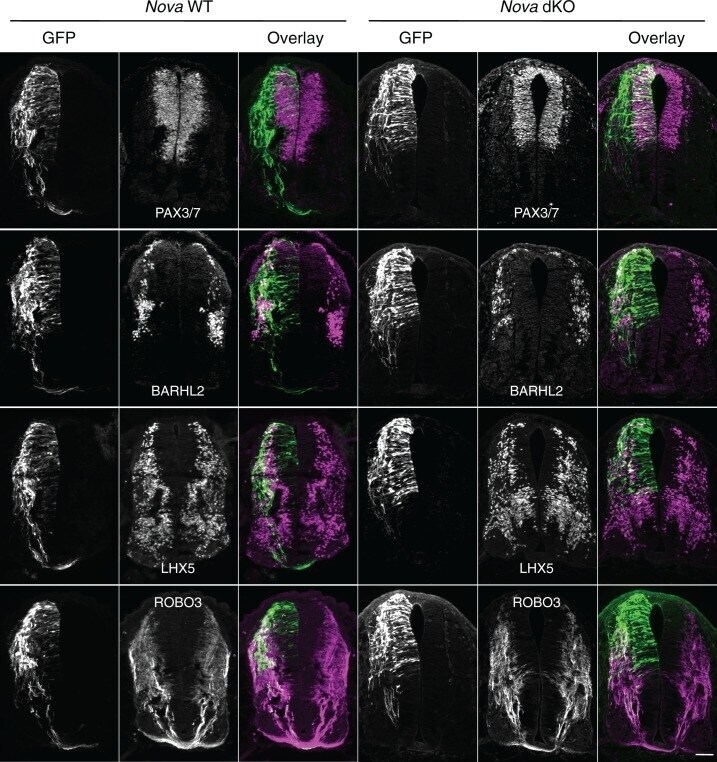
- Figure 2. Expression of neuronal and axonal markers in cultured Nova WT and dKO embryos electroporated with Actb-gfp . PAX3/7 immunostaining labels dorsal interneuron progenitors, which reside within the ventricular zone and give rise to different populations of interneurons. BARHL2 and LHX5 stainings label differentiated interneurons at the lateral spinal cord. ROBO3 staining specifically labels the commissural axons. Scale bar, 50 mum. DOI: http://dx.doi.org/ Figure 2--figure supplement 1. Fluorescent in situ hybridization of Dcc in cultured embryos electroporated with Actb-gfp . Nova dKO was used as there are more GFP+ neurons in the VZ. Dcc expression is seen in GFP+ neuroprogenitors and interneurons. Scale bars, 50 mum. DOI: http://dx.doi.org/
- Submitted by
- Invitrogen Antibodies (provider)
- Main image
- Experimental details
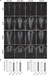
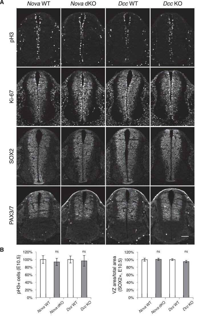
- Figure 3--figure supplement 1. Neural stem cells and progenitors appear normal in Nova and Dcc KOs. ( A ) Immunohistochemistry of phospho-histone H3, a mitotic marker, Ki-67, a cell proliferation marker, SOX2, a neural progenitor marker, and PAX3/7, a dorsal interneuron progenitor marker, in E10.5 spinal cords. ( B ) Quantification of phenotypes in A. pH3+ cells were counted and normalized to WT. The ratio between the SOX2+ VZ area and the total spinal cord area was quantified and normalized to WT. Data are represented as the mean +- SEM (Student's t-test, ns, not significant). Scale bar, 50 mum. DOI: http://dx.doi.org/
- Submitted by
- Invitrogen Antibodies (provider)
- Main image

- Experimental details
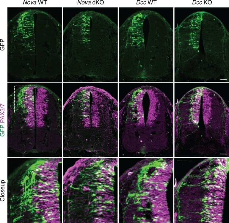
- Figure 3. Nova and Dcc knockouts delay the lateral migration of dorsal interneuron progenitors. Actb-gfp was electroporated into neural progenitors and embryos were cultured for 20 hrs. The bottom panel shows the closeup images of the boxed area from the middle panel. Many WT neurons have migrated out of the VZ (demarcated by PAX3/7 staining) after 20 hrs, whereas Nova and Dcc KO neurons are mostly located within the VZ. Scale bars, 50 mum. DOI: http://dx.doi.org/ Figure 3--figure supplement 1. Neural stem cells and progenitors appear normal in Nova and Dcc KOs. ( A ) Immunohistochemistry of phospho-histone H3, a mitotic marker, Ki-67, a cell proliferation marker, SOX2, a neural progenitor marker, and PAX3/7, a dorsal interneuron progenitor marker, in E10.5 spinal cords. ( B ) Quantification of phenotypes in A. pH3+ cells were counted and normalized to WT. The ratio between the SOX2+ VZ area and the total spinal cord area was quantified and normalized to WT. Data are represented as the mean +- SEM (Student's t-test, ns, not significant). Scale bar, 50 mum. DOI: http://dx.doi.org/ Figure 3--figure supplement 2. Dorsal interneuron differentiation is normal in Nova and Dcc KOs. ( A ) Immunohistochemistry of ISL1/2 and LHX5 in Nova WT, Nova dKO, Dcc WT, and Dcc KO spinal cords at E10.5. The markers are expressed by different subpopulations of interneurons in the dorsal spinal cord. ( B ) Quantification of ISL1/2+ neurons located in the dorsal half of the spinal cord. Data ar