Explore
 Validate
 Learn
Learn38-1801
antibody from Invitrogen Antibodies
Targeting: PAX3
HUP2, WS1
 Western blot
 Immunocytochemistry
 Immunoprecipitation Immunohistochemistry Flow cytometry Other assay
Antibody data
- Antibody Data
- Antigen structure
- References [10]
- Comments [0]
- Validations
- Immunocytochemistry [2]
- Immunohistochemistry [2]
- Flow cytometry [1]
- Other assay [8]
Submit
Validation data
Reference
Comment
Report error
- Product number
- 38-1801 - Provider product page

- Provider
- Invitrogen Antibodies
- Product name
- PAX3 Polyclonal Antibody
- Antibody type
- Polyclonal
- Antigen
- Recombinant full-length protein
- Reactivity
- Human, Mouse
- Host
- Rabbit
- Isotype
- IgG
- Vial size
- 100 μL
- Concentration
- Conc. Not Determined
- Storage
- -20°C
Submitted references 3D Interfacial and Spatiotemporal Regulation of Human Neuroepithelial Organoids.
Therapeutic potential of mesenchymal stem cells for peripheral artery disease in a rat model of hindlimb ischemia.
The protective role of DOT1L in UV-induced melanomagenesis.
Loss of Ezh2 promotes a midbrain-to-forebrain identity switch by direct gene derepression and Wnt-dependent regulation.
Recurrent PAX3-MAML3 fusion in biphenotypic sinonasal sarcoma.
SOX2-LIN28/let-7 pathway regulates proliferation and neurogenesis in neural precursors.
Direct reprogramming of melanocytes to neural crest stem-like cells by one defined factor.
Pax3-FKHR knock-in mice show developmental aberrations but do not develop tumors.
The EF-hand calcium-binding protein calmyrin inhibits the transcriptional and DNA-binding activity of Pax3.
The oncogenic potential of the Pax3-FKHR fusion protein requires the Pax3 homeodomain recognition helix but not the Pax3 paired-box DNA binding domain.
Tang C, Wang X, D'Urso M, van der Putten C, Kurniawan NA
Advanced science (Weinheim, Baden-Wurttemberg, Germany) 2022 Aug;9(22):e2201106
Advanced science (Weinheim, Baden-Wurttemberg, Germany) 2022 Aug;9(22):e2201106
Therapeutic potential of mesenchymal stem cells for peripheral artery disease in a rat model of hindlimb ischemia.
Ali AMEA, Ahmed AS, El-Yasergy DF, Abousarie MA, Elsayed RM, Mohammed YE, Mohammed RA
Iranian journal of basic medical sciences 2021 Jun;24(6):805-814
Iranian journal of basic medical sciences 2021 Jun;24(6):805-814
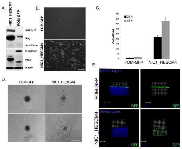
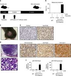
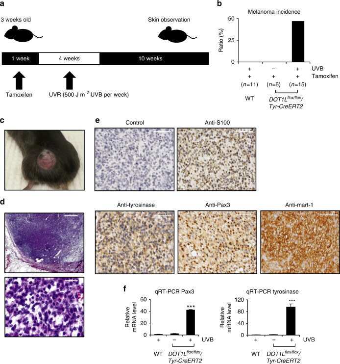
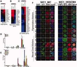
The protective role of DOT1L in UV-induced melanomagenesis.
Zhu B, Chen S, Wang H, Yin C, Han C, Peng C, Liu Z, Wan L, Zhang X, Zhang J, Lian CG, Ma P, Xu ZX, Prince S, Wang T, Gao X, Shi Y, Liu D, Liu M, Wei W, Wei Z, Pan J, Wang Y, Xuan Z, Hess J, Hayward NK, Goding CR, Chen X, Zhou J, Cui R
Nature communications 2018 Jan 17;9(1):259
Nature communications 2018 Jan 17;9(1):259
Loss of Ezh2 promotes a midbrain-to-forebrain identity switch by direct gene derepression and Wnt-dependent regulation.
Zemke M, Draganova K, Klug A, Schöler A, Zurkirchen L, Gay MH, Cheng P, Koseki H, Valenta T, Schübeler D, Basler K, Sommer L
BMC biology 2015 Nov 30;13:103
BMC biology 2015 Nov 30;13:103
Recurrent PAX3-MAML3 fusion in biphenotypic sinonasal sarcoma.
Wang X, Bledsoe KL, Graham RP, Asmann YW, Viswanatha DS, Lewis JE, Lewis JT, Chou MM, Yaszemski MJ, Jen J, Westendorf JJ, Oliveira AM
Nature genetics 2014 Jul;46(7):666-8
Nature genetics 2014 Jul;46(7):666-8
SOX2-LIN28/let-7 pathway regulates proliferation and neurogenesis in neural precursors.
Cimadamore F, Amador-Arjona A, Chen C, Huang CT, Terskikh AV
Proceedings of the National Academy of Sciences of the United States of America 2013 Aug 6;110(32):E3017-26
Proceedings of the National Academy of Sciences of the United States of America 2013 Aug 6;110(32):E3017-26
Direct reprogramming of melanocytes to neural crest stem-like cells by one defined factor.
Zabierowski SE, Baubet V, Himes B, Li L, Fukunaga-Kalabis M, Patel S, McDaid R, Guerra M, Gimotty P, Dahmane N, Herlyn M
Stem cells (Dayton, Ohio) 2011 Nov;29(11):1752-62
Stem cells (Dayton, Ohio) 2011 Nov;29(11):1752-62
Pax3-FKHR knock-in mice show developmental aberrations but do not develop tumors.
Lagutina I, Conway SJ, Sublett J, Grosveld GC
Molecular and cellular biology 2002 Oct;22(20):7204-16
Molecular and cellular biology 2002 Oct;22(20):7204-16
The EF-hand calcium-binding protein calmyrin inhibits the transcriptional and DNA-binding activity of Pax3.
Hollenbach AD, McPherson CJ, Lagutina I, Grosveld G
Biochimica et biophysica acta 2002 Apr 12;1574(3):321-8
Biochimica et biophysica acta 2002 Apr 12;1574(3):321-8
The oncogenic potential of the Pax3-FKHR fusion protein requires the Pax3 homeodomain recognition helix but not the Pax3 paired-box DNA binding domain.
Lam PY, Sublett JE, Hollenbach AD, Roussel MF
Molecular and cellular biology 1999 Jan;19(1):594-601
Molecular and cellular biology 1999 Jan;19(1):594-601
No comments: Submit comment
Supportive validation
- Submitted by
- Invitrogen Antibodies (provider)
- Main image

- Experimental details
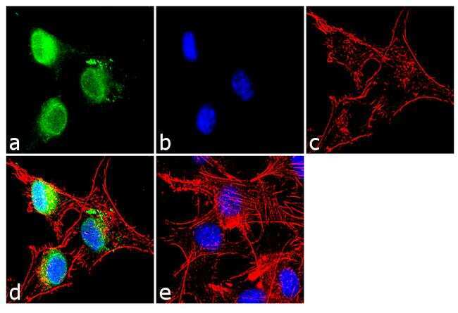
- Immunofluorescence analysis of PAX3 was performed using 70% confluent log phase U-87 MG cells. The cells were fixed with 4% paraformaldehyde for 10 minutes, permeabilized with 0.1% Triton™ X-100 for 10 minutes, and blocked with 2% BSA for 1 hour at room temperature. The cells were labeled with PAX3 Rabbit Polyclonal Antibody (Product # 38-1801) at 1:250 dilution in 0.1% BSA and incubated for 3 hours at room temperature and then labeled with Goat anti-Rabbit IgG (H+L) Superclonal™ Secondary Antibody, Alexa Fluor® 488 conjugate (Product # A27034) a dilution of 1:2000 for 45 minutes at room temperature (Panel a: green). Nuclei (Panel b: blue) were stained with SlowFade® Gold Antifade Mountant with DAPI (Product # S36938). F-actin (Panel c: red) was stained with Rhodamine Phalloidin (Product # R415, 1:300). Panel d represents the merged image showing nuclear localization. Panel e shows the no primary antibody control. The images were captured at 60X magnification.
- Submitted by
- Invitrogen Antibodies (provider)
- Main image
- Experimental details
- Immunofluorescence analysis of PAX3 was performed using 70% confluent log phase U-87 MG cells. The cells were fixed with 4% paraformaldehyde for 10 minutes, permeabilized with 0.1% Triton™ X-100 for 10 minutes, and blocked with 2% BSA for 1 hour at room temperature. The cells were labeled with PAX3 Rabbit Polyclonal Antibody (Product # 38-1801) at 1:250 dilution in 0.1% BSA and incubated for 3 hours at room temperature and then labeled with Goat anti-Rabbit IgG (Heavy Chain) Superclonal™ Secondary Antibody, Alexa Fluor® 488 conjugate (Product # A27034) a dilution of 1:2000 for 45 minutes at room temperature (Panel a: green). Nuclei (Panel b: blue) were stained with SlowFade® Gold Antifade Mountant with DAPI (Product # S36938). F-actin (Panel c: red) was stained with Rhodamine Phalloidin (Product # R415, 1:300). Panel d represents the merged image showing nuclear localization. Panel e shows the no primary antibody control. The images were captured at 60X magnification.

Supportive validation
- Submitted by
- Invitrogen Antibodies (provider)
- Main image
- Experimental details
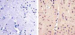
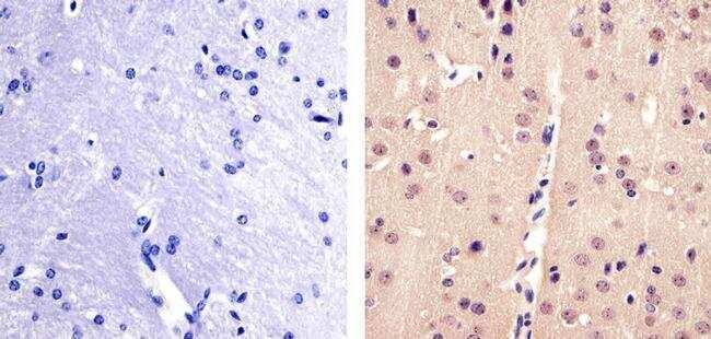
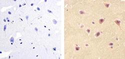
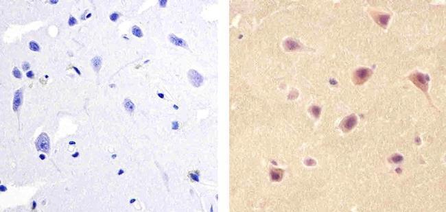
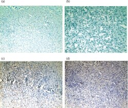
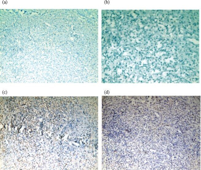
- Immunohistochemistry analysis of PAX3 showing staining in the nucleus of paraffin-embedded mouse brain tissue (right) compared to a negative control without primary antibody (left). To expose target proteins, antigen retrieval was performed using 10mM sodium citrate (pH 6.0), microwaved for 8-15 min. Following antigen retrieval, tissues were blocked in 3% H2O2-methanol for 15 min at room temperature, washed with ddH2O and PBS, and then probed with a Anti- PAX3 Polyclonal Antibody (Product # 38-1801) diluted in 3% BSA-PBS at a dilution of 1:100 overnight at 4°C in a humidified chamber. Tissues were washed extensively in PBST and detection was performed using an HRP-conjugated secondary antibody followed by colorimetric detection using a DAB kit. Tissues were counterstained with hematoxylin and dehydrated with ethanol and xylene to prep for mounting.
- Submitted by
- Invitrogen Antibodies (provider)
- Main image
- Experimental details
- Immunohistochemistry analysis of PAX3 showing staining in the nucleus of paraffin-embedded human brain tissue (right) compared to a negative control without primary antibody (left). To expose target proteins, antigen retrieval was performed using 10mM sodium citrate (pH 6.0), microwaved for 8-15 min. Following antigen retrieval, tissues were blocked in 3% H2O2-methanol for 15 min at room temperature, washed with ddH2O and PBS, and then probed with a Anti- PAX3 Polyclonal Antibody (Product # 38-1801) diluted in 3% BSA-PBS at a dilution of 1:100 overnight at 4°C in a humidified chamber. Tissues were washed extensively in PBST and detection was performed using an HRP-conjugated secondary antibody followed by colorimetric detection using a DAB kit. Tissues were counterstained with hematoxylin and dehydrated with ethanol and xylene to prep for mounting.
Supportive validation
- Submitted by
- Invitrogen Antibodies (provider)
- Main image
- Experimental details
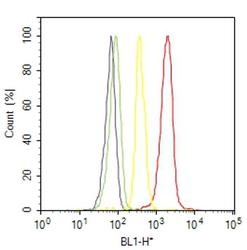
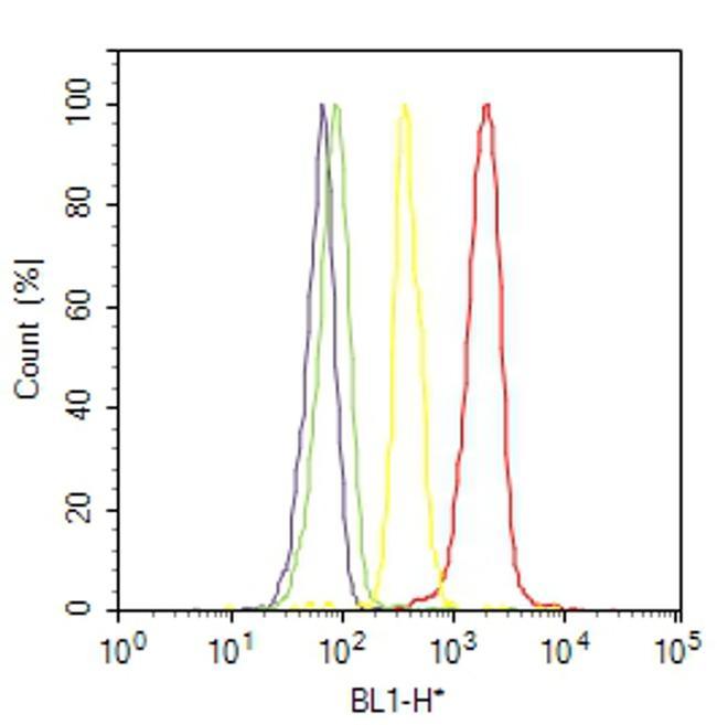
- Flow cytometry analysis of PAX3 was done on U-87 MG. Cells were fixed with 70% ethanol for 10 minutes, permeabilized with 0.25% Triton™ X-100 for 20 minutes, and blocked with 5% BSA for 30 minutes at room temperature. Cells were labeled with PAX3 Rabbit Polyclonal Antibody (381801, red histogram) or with rabbit isotype control (yellow histogram) at 3-5 ug/million cells in 2.5% BSA. After incubation at room temperature for 2 hours, the cells were labeled with Alexa Fluor® 488 Goat Anti-Rabbit Secondary Antibody (A11008) at a dilution of 1:400 for 30 minutes at room temperature. The representative 10,000 cells were acquired and analyzed for each sample using an Attune® Acoustic Focusing Cytometer. The purple histogram represents unstained control cells and the green histogram represents no-primary-antibody control.
Supportive validation
- Submitted by
- Invitrogen Antibodies (provider)
- Main image
- Experimental details
- NULL
- Submitted by
- Invitrogen Antibodies (provider)
- Main image
- Experimental details
- NULL
- Submitted by
- Invitrogen Antibodies (provider)
- Main image
- Experimental details
- NULL
- Submitted by
- Invitrogen Antibodies (provider)
- Main image
- Experimental details
- NULL
- Submitted by
- Invitrogen Antibodies (provider)
- Main image
- Experimental details
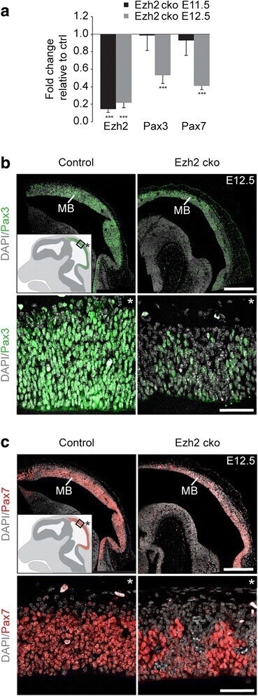
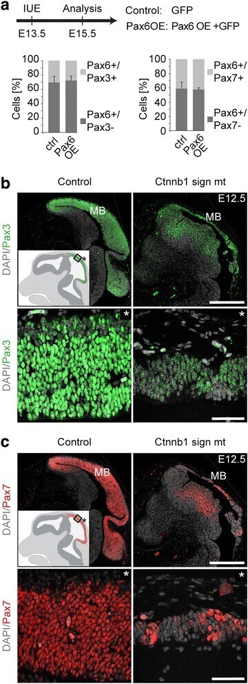
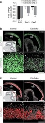
- Fig. 6 Midbrain markers are not directly repressed by Pax6 but are regulated indirectly by Wnt/beta-catenin signaling. ( a ) A Pax6 overexpression construct together with a GFP-expressing vector (Pax6OE) or a GFP-expressing vector alone (Control) were delivered by in utero electroporation into the dorsal midbrain at E13.5. Two days later the proportion of GFP + cells expressing midbrain markers Pax3 and Pax7 was analyzed. Overexpression of Pax6 does not affect the proportion of midbrain marker-expressing cells. n = 3 in each group, two different litters (also see Additional file 1 : Figure S7B). ( b , c ) Immunostaining for Pax3 ( b ) and Pax7 ( c ) at E12.5 reveals decreased protein levels in midbrains with ablated Wnt/beta-catenin signaling (Ctnnb1 sign mt) compared to control. Cartoon insets show regular expression pattern, asterisk indicates basal. DAPI staining serves as nuclear marker: b , c ; Scale bars: b , c , 500 mum (upper panel); b , c , 40 mum (lower panel); Error bars indicate SD; IUE, in utero electroporation; ctrl, control; Ctnnb1 sign mt, Wnt/beta-catenin signaling mutant; MB, midbrain
- Submitted by
- Invitrogen Antibodies (provider)
- Main image
- Experimental details
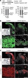
- Figure 5 Photomicrograph of muscle sections in ischemic non-treated and ischemic treated groups by BM-MSCs stained for Pax3 and Pax7 nuclear expression. (a) The granulation tissue of the ischemic non-treated group showing negative expression of Pax3 (diaminobenzidine, original magnification, x200). (b) The granulation tissue of the ischemic non-treated group showing negative expression of Pax7 (diaminobenzidine, original magnification, x400). (c) The granulation tissue of the ischemic treated group by BM-MSCs showing cells with positive nuclear staining of Pax3 (diaminobenzidine, original magnification, x200). (d) The granulation tissue of the ischemic treated group by BM-MSCs showing cells with positive nuclear staining of Pax7 (diaminobenzidine, original magnification, x200)
- Submitted by
- Invitrogen Antibodies (provider)
- Main image
- Experimental details
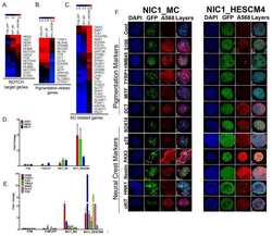
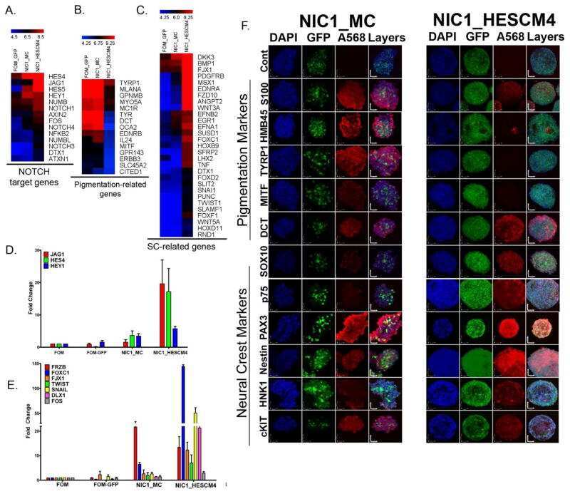
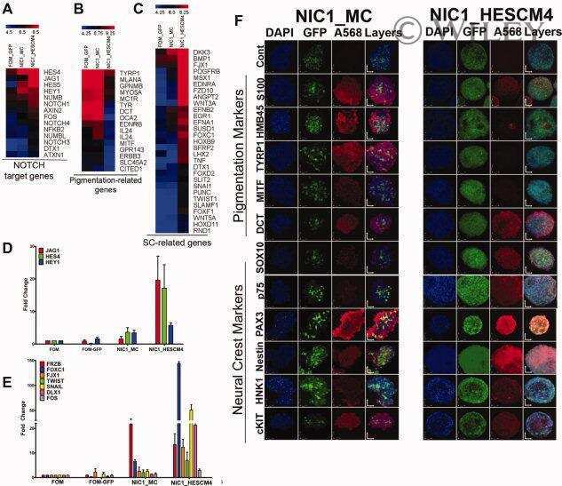
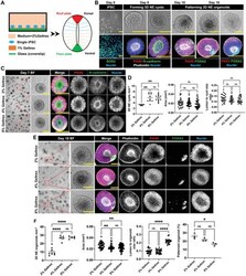
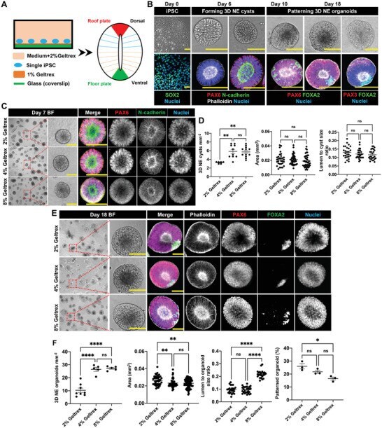
- Formation of 3D NE organoids from human iPSCs. A) Culture system for generating 3D DV patterned NE organoids from single hiPSCs. B) Developmental phases during the formation of NE organoids from single hiPSCs. Phase 1, hiPSCs were digested into single cells and seeded on Geltrex-coated glass coverslip on day 0. Phase 2, hiPSCs differentiated into NE cells and self-organized into cystic structures with smooth outer surface and containing central lumen with apical-basal polarity on day 6. Phase 3, development of NE cysts with FOXA2 + ventral NE cells and PAX3 + dorsal NE cells, forming DV patterned NE organoids. Top panels show bright-field images of hiPSCs, NE cysts, and NE organoids. Bottom panels show immunofluorescence staining of pluripotency marker SOX2, F-actin (phalloidin), N-cadherin, early neuroectodermal marker PAX6, dorsal marker PAX3, and ventral marker FOXA2 in corresponding phases during NE organoid formation. Scale bars: 100 um. C) Representative bright-field (BF) images of NE cysts on day 7 and the corresponding immunofluorescence staining of PAX6, N-cadherin, and nuclei in NE cysts formed in the presence of 2%, 4%, and 8% Geltrex in the neural induction medium. Scale bars: 100 um. D) Quantification of the number density, area, and lumen-to-cyst size ratio of NE cysts on day 7, n >= 5. E) Representative bright-field (BF) images (scale bars: 100 um) and the corresponding immunofluorescence staining (scale bars: 50 um) of F-actin (phalloidin), PAX6, FOXA2, and nu
- Submitted by
- Invitrogen Antibodies (provider)
- Main image
- Experimental details
- Fig. 5 Ezh2 regulates midbrain identity indirectly. ( a ) qRT-PCR for Ezh2 , Pax3 , and Pax7 on midbrain tissue isolated at E11.5 and E12.5 reveals a downregulation of midbrain transcription factors Pax3 and Pax7 in the absence of Ezh2 after E11.5. n >=3 in each group, *** P