Explore
 Validate
 Learn
Learn Western blot
 ELISA
ELISAAntibody data
- Antibody Data
- Antigen structure
- References [1]
- Comments [0]
- Validations
- Western blot [1]
Submit
Validation data
Reference
Comment
Report error
- Product number
- A00285-1 - Provider product page

- Provider
- Boster Biological Technology
- Product name
- Anti-PAX3 Antibody Picoband™
- Antibody type
- Polyclonal
- Description
- Rabbit IgG polyclonal antibody for PAX3 detection. Tested with WB, FCM, Direct ELISA in Human;Mouse;Rat.
- Reactivity
- Human, Mouse, Rat
- Host
- Rabbit
- Vial size
- 100μg/vial
- Concentration
- Add 0.2ml of distilled water will yield a concentration of 500ug/ml.
- Storage
- At -20°C for one year. After reconstitution, at 4°C for one month. It can also be aliquoted and stored frozen at -20°C for a longer time. Avoid repeated freezing and thawing.
- Handling
- Add 0.2ml of distilled water will yield a concentration of 500ug/ml.
Submitted references Silencing Pax3 by shRNA inhibits the proliferation and differentiation of duck (Anas platyrhynchos) myoblasts.
Zhang RP, Liu HH, Wang HH, Wang Y, Han CC, Li L, He H, Xu HY, Xu F, Wang JW
Molecular and cellular biochemistry 2014 Jan;386(1-2):211-22
Molecular and cellular biochemistry 2014 Jan;386(1-2):211-22
No comments: Submit comment
Supportive validation
- Submitted by
- Boster Biological Technology (provider)
- Main image
- Experimental details
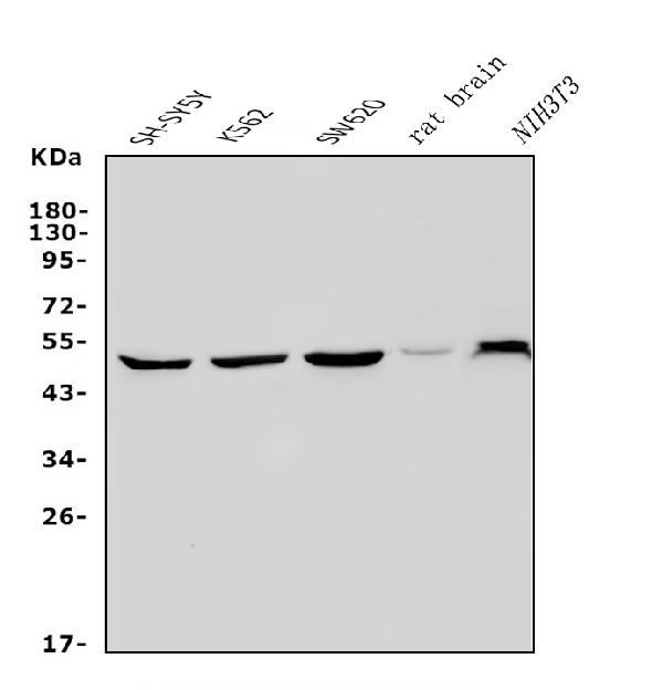
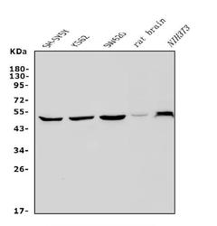
- Western blot analysis of PAX3 using anti-PAX3 antibody (A00285-1). Electrophoresis was performed on a 5-20% SDS-PAGE gel at 70V (Stacking gel) / 90V (Resolving gel) for 2-3 hours. The sample well of each lane was loaded with 30ug of sample under reducing conditions. Lane 1: human SH-SY5Y whole cell lysates, Lane 2: human K562 whole cell lysates, Lane 3: human SW620 whole cell lysates, Lane 4: rat brain tissue lysates, Lane 5: mouse NIH/3T3 whole cell lysates. After Electrophoresis, proteins were transferred to a Nitrocellulose membrane at 150mA for 50-90 minutes. Blocked the membrane with 5% Non-fat Milk/ TBS for 1.5 hour at RT. The membrane was incubated with rabbit anti-PAX3 antigen affinity purified polyclonal antibody (Catalog # A00285-1) at 0.5 μg/mL overnight at 4°C, then washed with TBS-0.1%Tween 3 times with 5 minutes each and probed with a goat anti-rabbit IgG-HRP secondary antibody at a dilution of 1:5000 for 1.5 hour at RT. The signal is developed using an Enhanced Chemiluminescent detection (ECL) kit (Catalog # EK1002) with Tanon 5200 system. A specific band was detected for PAX3 at approximately 53KD. The expected band size for PAX3 is at 53KD.
- Additional image