Explore
 Validate
 Learn
Learn Western blot
 Immunocytochemistry
Antibody data
- Antibody Data
- Antigen structure
- References [2]
- Comments [0]
- Validations
- Immunocytochemistry [3]
- Immunohistochemistry [1]
- Other assay [4]
Submit
Validation data
Reference
Comment
Report error
- Product number
- PA5-21599 - Provider product page

- Provider
- Invitrogen Antibodies
- Product name
- SOCS4 Polyclonal Antibody
- Antibody type
- Polyclonal
- Antigen
- Synthetic peptide
- Description
- Recommended positive controls: HeLaS3. Predicted reactivity: Pig (100%), Rhesus Monkey (100%), Bovine (100%). Store product as a concentrated solution. Centrifuge briefly prior to opening the vial.
- Reactivity
- Human
- Host
- Rabbit
- Isotype
- IgG
- Vial size
- 100 μL
- Concentration
- 1 mg/mL
- Storage
- Store at 4°C short term. For long term storage, store at -20°C, avoiding freeze/thaw cycles.
Submitted references Up-regulation of SOCS4 promotes cell proliferation and migration in esophageal squamous cell carcinoma.
Maintenance of cancer stemness by miR-196b-5p contributes to chemoresistance of colorectal cancer cells via activating STAT3 signaling pathway.
Ying J, Huang HH, Zhang MM, Chen JF
Translational cancer research 2021 May;10(5):2416-2427
Translational cancer research 2021 May;10(5):2416-2427
Maintenance of cancer stemness by miR-196b-5p contributes to chemoresistance of colorectal cancer cells via activating STAT3 signaling pathway.
Ren D, Lin B, Zhang X, Peng Y, Ye Z, Ma Y, Liang Y, Cao L, Li X, Li R, Sun L, Liu Q, Wu J, Zhou K, Zeng J
Oncotarget 2017 Jul 25;8(30):49807-49823
Oncotarget 2017 Jul 25;8(30):49807-49823
No comments: Submit comment
Supportive validation
- Submitted by
- Invitrogen Antibodies (provider)
- Main image

- Experimental details
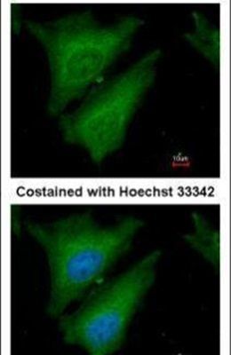
- Immunofluorescent analysis of SOCS4 in paraformaldehyde-fixed HeLa cells using a SOCS4 polyclonal antibody (Product # PA5-21599) at a 1:200 dilution.
- Submitted by
- Invitrogen Antibodies (provider)
- Main image

- Experimental details
- Immunofluorescence analysis of paraformaldehyde-fixed HeLa, using SOCS4 (Product # PA5-21599) antibody at 1:200 dilution.

- Submitted by
- Invitrogen Antibodies (provider)
- Main image

- Experimental details
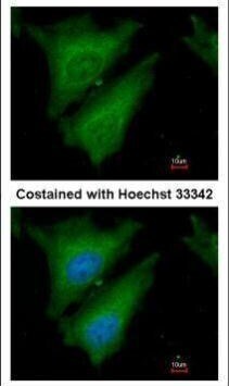
- Immunofluorescence analysis of paraformaldehyde-fixed HeLa, using SOCS4 (Product # PA5-21599) antibody at 1:200 dilution.
Supportive validation
- Submitted by
- Invitrogen Antibodies (provider)
- Main image
- Experimental details
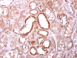
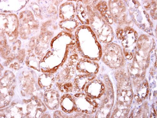
- SOCS4 Polyclonal Antibody detects SOCS4 protein at cytoplasm on human normal kidney by immunohistochemical analysis. Sample: Paraffin-embedded human normal kidney. SOCS4 Polyclonal Antibody (Product # PA5-21599) diluted at 1:500. Antigen Retrieval: EDTA based buffer, pH 8.0, 15 min.
Supportive validation
- Submitted by
- Invitrogen Antibodies (provider)
- Main image

- Experimental details
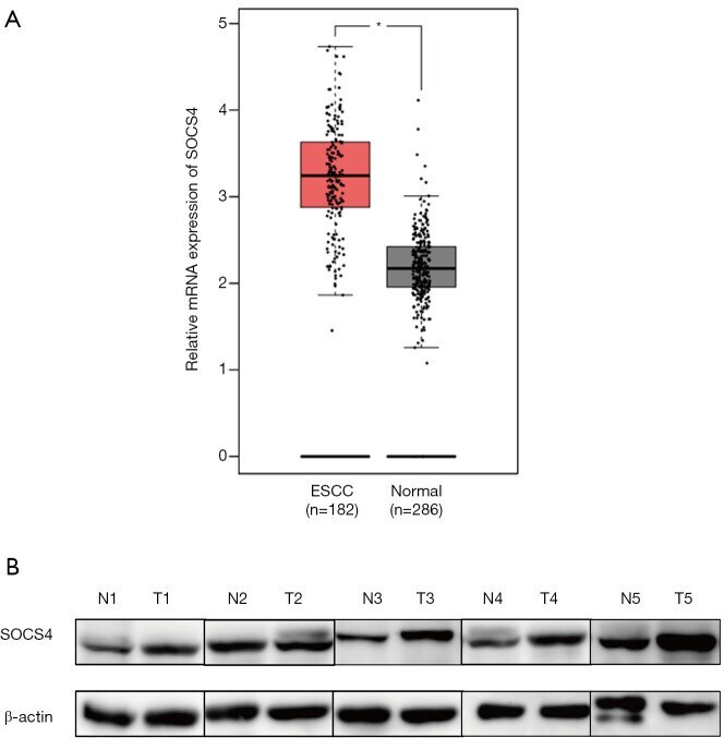
- Figure 1 SOCS4 expression was up-regulated in ESCC tissues. SOCS4 mRNA (A) expressions in ESCC tissue and normal tissues in TCGA database (A), SOCS4 protein expressions in ESCC tissue (T) and normal tissues (N) were measured using western blotting (B). *, P
- Submitted by
- Invitrogen Antibodies (provider)
- Main image
- Experimental details
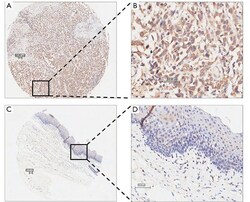
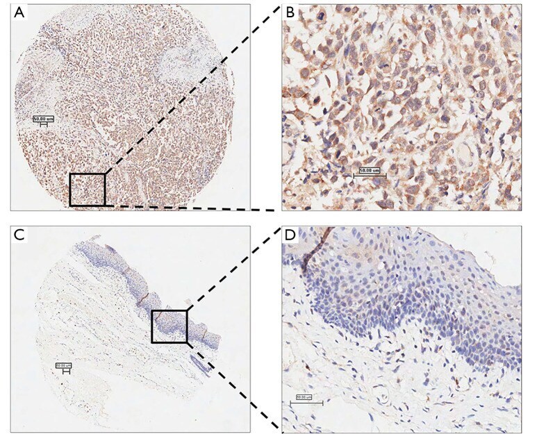
- Figure 2 SOCS4 expression in ESCC tissues and adjacent normal tissues by immunohistochemistry. Representative immunostaining of SOCS4 in human ESCC tissues (A,B) and adjacent normal tissues (C,D), magnification 40x (A,C), magnification 200x (B,D). SOCS4, suppressors of cytokine signaling family member 4; ESCC, esophageal squamous cell carcinoma, Scale bar: 50 um.
- Submitted by
- Invitrogen Antibodies (provider)
- Main image
- Experimental details
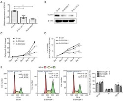
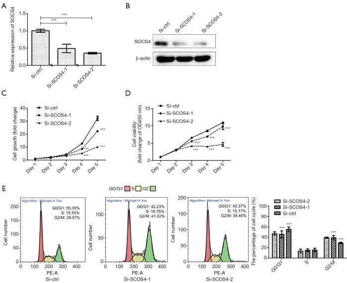
- Figure 4 SOCS4 knockdown inhibits ESCC cell proliferation. SOCS4 expression in Eca-109 cells following transfection with control siRNA (Si-ctrl), SOCS4 siRNAs (Si-SOCS4-1 and Si-SOCS4-1) were measured by RT-qPCR (A) and western blot (B), respectively. The effect of SOCS4 knockdown on cell proliferation. Eca-109 cells transiently transfected with Si-ctrl, Si-SOCS4-1, and Si-SOCS4-1 were cultured for 72 h. Cell growth was measured by counting cell numbers (C,D), and cell viability was measured using an CCK-8 assay. The effect of SOCS4 knockdown on cell cycle was measured by flow cytometry (E). ***, P
- Submitted by
- Invitrogen Antibodies (provider)
- Main image
- Experimental details
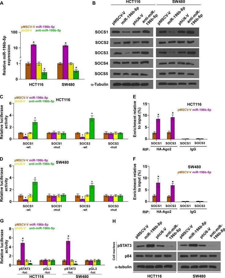
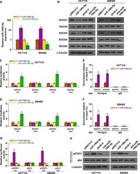
- Figure 2 miR-196b-5p activates STAT3 signaling via targeting multiple negative regulators of STAT3 signaling ( A ) Real-time PCR analysis of miR-196b-5p expression in the indicated cells. Transcript levels were normalized by U6 expression. Error bars represent the mean +- SD of three independent experiments. * P < 0.05. ( B ) Western blotting of SOCS1, SOCS2, SOCS3, SOCS4 and SOCS5 expression in the indicated cells. alpha-Tubulin served as the loading control. ( C and D ) Luciferase assay of cells transfected with pmirGLO-3'UTR reporter of SOCS1 and SOCS3 in miR-196b-5p overexpressing and silencing HCT116 and SW480 cells, respectively. Error bars represent the mean +- SD of three independent experiments. * P < 0.05. ( E and F ) MiRNP IP assay showing the association between miR-196b-5p and SOCS1,SOCS3 transcripts in HCT116 and SW480 cells. Pulldown of IgGantibody served as the negative control. Error bars represent the mean +- SD of three independent experiments. * P < 0.05. ( G ) STAT3 transcriptional activity was assessed by luciferase reporter constructs in the indicated cells. Error bars represent the mean +- SD of three independent experiments. * P < 0.05. ( H ) Western blotting of nuclear STAT3 expression. The nuclear protein p84 was used as the nuclear protein marker.