 Explore
 Validate
 Learn
Learn Western blot
 ELISA
ELISAAntibody data
- Antibody Data
- Antigen structure
- References [3]
- Comments [0]
- Validations
- Western blot [1]
- Immunocytochemistry [1]
Submit
Validation data
Reference
Comment
Report error
- Product number
- PAB10250 - Provider product page

- Provider
- Abnova Corporation
- Proper citation
- Abnova Corporation Cat#PAB10250, RRID:AB_1676577
- Product name
- IRAK2 polyclonal antibody
- Antibody type
- Polyclonal
- Antigen
- A synthetic peptide corresponding to C-terminus of human IRAK2.
- Reactivity
- Human
- Host
- Rabbit
- Vial size
- 100 μg
- Storage
- Store at 4°C. For long term storage store at -20°C.Aliquot to avoid repeated freezing and thawing.
Submitted references Bacterial lipopolysaccharide activates nuclear factor-kappaB through interleukin-1 signaling mediators in cultured human dermal endothelial cells and mononuclear phagocytes.
Signaling events induced by lipopolysaccharide-activated toll-like receptor 2.
IRAK (Pelle) family member IRAK-2 and MyD88 as proximal mediators of IL-1 signaling.
Zhang FX, Kirschning CJ, Mancinelli R, Xu XP, Jin Y, Faure E, Mantovani A, Rothe M, Muzio M, Arditi M
The Journal of biological chemistry 1999 Mar 19;274(12):7611-4
The Journal of biological chemistry 1999 Mar 19;274(12):7611-4
Signaling events induced by lipopolysaccharide-activated toll-like receptor 2.
Yang RB, Mark MR, Gurney AL, Godowski PJ
Journal of immunology (Baltimore, Md. : 1950) 1999 Jul 15;163(2):639-43
Journal of immunology (Baltimore, Md. : 1950) 1999 Jul 15;163(2):639-43
IRAK (Pelle) family member IRAK-2 and MyD88 as proximal mediators of IL-1 signaling.
Muzio M, Ni J, Feng P, Dixit VM
Science (New York, N.Y.) 1997 Nov 28;278(5343):1612-5
Science (New York, N.Y.) 1997 Nov 28;278(5343):1612-5
No comments: Submit comment
Supportive validation
- Submitted by
- Abnova Corporation (provider)
- Main image
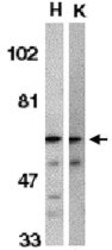
- Experimental details
- Western blot using IRAK2 polyclonal antibody (Cat # PAB10250) shows detection of IRAK2 in HeLa (H) and K-562 (K) whole cell lysates. The membrane was probed with the primary antibody diluted to 1 : 500.

Supportive validation
- Submitted by
- Abnova Corporation (provider)
- Main image
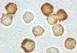
- Experimental details
- Immunocytochemistry using IRAK2 polyclonal antibody (Cat # PAB10250) shows staining of IRAK2 in HeLa cells.The cells were fixed with paraformaldehyde and reacted with the primary antibody diluted to 1 : 500.
- Validation comment
- Immunocytochemistry
