Explore
 Validate
 Learn
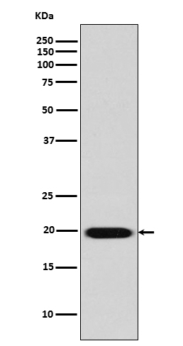
Learn Western blot
 Immunohistochemistry
Antibody data
- Antibody Data
- Antigen structure
- References [0]
- Comments [0]
- Validations
- Immunohistochemistry [1]
Submit
Validation data
Reference
Comment
Report error
- Product number
- M01574 - Provider product page

- Provider
- Boster Biological Technology
- Product name
- Anti-Caveolin-2 CAV2 Rabbit Monoclonal Antibody
- Antibody type
- Monoclonal
- Description
- Monoclonal antibody for CAVEOLIN 2/CAV2 detection. Host: Rabbit.Size: 100ug/vial. Tested applications: IP, IF, IHC, ICC, WB. Reactive species: Human CAVEOLIN 2/CAV2 information: Molecular Weight: 18291 MW; Subcellular Localization: Nucleus. Cytoplasm. Golgi apparatus membrane; Peripheral membrane protein. Cell membrane; Peripheral membrane protein. Membrane, caveola; Peripheral membrane protein. Potential hairpin-like structure in the membrane. Membrane protein of caveolae. Tyr-19-phosphorylated form is enriched at sites of cell-cell contact and is translocated to the nucleus in complex with MAPK1 in response to insulin (By similarity). Tyr-27- phosphorylated form is located both in the cytoplasm and plasma membrane. CAV1-mediated Ser-23-phosphorylated form locates to the plasma membrane. Ser-36-phosphorylated form resides in intracellular compartments; Tissue Specificity: Expressed in endothelial cells, smooth muscle cells, skeletal myoblasts and fibroblasts.
- Reactivity
- Human
- Host
- Rabbit
- Antibody clone number
- FD-3
- Vial size
- 100ug/vial
- Concentration
- 0.5-1mg/ml, actual concentration vary by lot. Use suggested dilution ratio to decide dilution procedure.
- Storage
- At -20°C for one year. Avoid repeated freezing and thawing.
No comments: Submit comment
Supportive validation
- Submitted by
- Boster Biological Technology (provider)
- Main image
- Experimental details
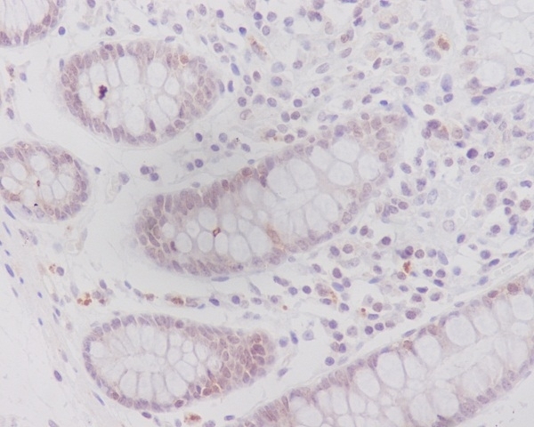
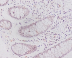
- Immunohistochemical analysis of paraffin-embedded human colon, using Caveolin-2 Antibody(M01574)CAV2 was detected in paraffin-embedded tissue section. Heat mediated antigen retrieval was performed in citrate buffer (pH6, epitope retrieval solution) for 20 mins. The tissue section was blocked with 10% goat serum. The tissue section was then incubated with 1ug/ml rabbit anti-CAV2 Antibody (M01574)overnight at 4?? Biotinylated goat anti-rabbit IgG was used as secondary antibody and incubated for 30 minutes at 37?? The tissue section was developed using Strepavidin-Biotin-Complex (SABC)(Catalog # SA1022) with DAB as the chromogen.
- Additional image