 Explore
 Validate
 Learn
Learn Western blot
 Immunocytochemistry
Antibody data
- Antibody Data
- Antigen structure
- References [2]
- Comments [0]
- Validations
- Immunocytochemistry [2]
- Other assay [1]
Submit
Validation data
Reference
Comment
Report error
- Product number
- PA1-060 - Provider product page

- Provider
- Invitrogen Antibodies
- Product name
- Phospho-Caveolin 2 (Tyr19) Polyclonal Antibody
- Antibody type
- Polyclonal
- Antigen
- Synthetic peptide
- Description
- PA1-060 detects phospho-caveolin-2 Y19 from human and mouse samples. PA1-060 has been successfully used in Western blot and immunofluorescence procedures. By Western blot, this antibody detects an ~21 kDa protein representing phospho-caveolin-2 Y19 from extract from COS-7 cells transiently transfected with the mouse caveolin-2 gene. Immunofluorescent staining of phospho-caveolin-2 Y19 in NIH 3T3 cells using PA1-060 results in a distinct punctate staining pattern at the cell periphery. The PA1-060 immunogen is a synthetic phosphopeptide corresponding to residues M(14) A D D A (pY) S H H S G C(25) of mouse CAV2. This peptide (Cat. # PEP-180) is available for use in neutralization and control experiments.
- Reactivity
- Human, Mouse
- Host
- Rabbit
- Isotype
- IgG
- Vial size
- 100 µg
- Concentration
- 1 mg/mL
- Storage
- -20° C, Avoid Freeze/Thaw Cycles
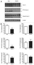
Submitted references Genetic ablation of caveolin-2 sensitizes mice to bleomycin-induced injury.
Src-induced phosphorylation of caveolin-2 on tyrosine 19. Phospho-caveolin-2 (Tyr(P)19) is localized near focal adhesions, remains associated with lipid rafts/caveolae, but no longer forms a high molecular mass hetero-oligomer with caveolin-1.
de Almeida CJ, Jasmin JF, Del Galdo F, Lisanti MP
Cell cycle (Georgetown, Tex.) 2013 Jul 15;12(14):2248-54
Cell cycle (Georgetown, Tex.) 2013 Jul 15;12(14):2248-54
Src-induced phosphorylation of caveolin-2 on tyrosine 19. Phospho-caveolin-2 (Tyr(P)19) is localized near focal adhesions, remains associated with lipid rafts/caveolae, but no longer forms a high molecular mass hetero-oligomer with caveolin-1.
Lee H, Park DS, Wang XB, Scherer PE, Schwartz PE, Lisanti MP
The Journal of biological chemistry 2002 Sep 13;277(37):34556-67
The Journal of biological chemistry 2002 Sep 13;277(37):34556-67
No comments: Submit comment
Supportive validation
- Submitted by
- Invitrogen Antibodies (provider)
- Main image
- Experimental details
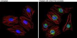
- Immunofluorescent analysis of Phospho-Caveolin 2 pTyr19 (green) showing staining in the cytoplasm and nucleus of HUVEC cells treated with 100µM pervanadate (left) and untreated HUVEC cells (right). Formalin-fixed cells were permeabilized with 0.1% Triton X-100 in TBS for 5-10 minutes and blocked with 3% BSA-PBS for 30 minutes at room temperature. Cells were probed with a Phospho-Caveolin 2 pTyr19 polyclonal antibody (Product # PA1-060) in 3% BSA-PBS at a dilution of 1:20 and incubated overnight at 4°C in a humidified chamber. Cells were washed with PBST and incubated with a DyLight-conjugated secondary antibody in PBS at room temperature in the dark. F-actin (red) was stained with a fluorescent red phalloidin and nuclei (blue) were stained with Hoechst or DAPI. Images were taken at a magnification of 60x.
- Submitted by
- Invitrogen Antibodies (provider)
- Main image
- Experimental details
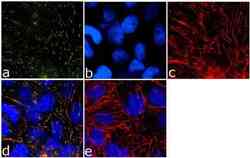
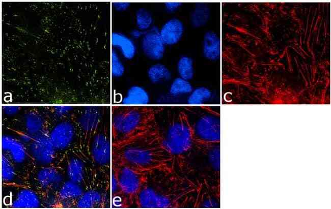
- Immunofluorescence analysis of Phospho-Caveolin 2 pTyr19 was done on 70% confluent log phase A-375 cells. The cells were fixed with 4% paraformaldehyde for 15 minutes, permeabilized with 0.25% Triton™ X-100 for 10 minutes, and blocked with 5% BSA for 1 hour at room temperature. The cells were labeled with Phospho-Caveolin 2 pTyr19 Rabbit Polyclonal Antibody (Product # PA1-060) at 1 µg/mL in 1% BSA and incubated for 3 hours at room temperature and then labeled with Goat anti-Rabbit IgG (H+L) Superclonal™ Secondary Antibody, Alexa Fluor® 488 conjugate (Product # A27034) at a dilution of 1:2000 for 45 minutes at room temperature (Panel a: green). Nuclei (Panel b: blue) were stained with SlowFade® Gold Antifade Mountant with DAPI (Product # S36938). F-actin (Panel c: red) was stained with Alexa Fluor® 555 Rhodamine Phalloidin (Product # R415, 1:300). Panel d is a merged image showing membranous localization. Panel e is a no primary antibody control. The images were captured at 60X magnification.
Supportive validation
- Submitted by
- Invitrogen Antibodies (provider)
- Main image
- Experimental details
- NULL
