Explore
 Validate
 Learn
Learn Western blot
 Immunocytochemistry
Antibody data
- Antibody Data
- Antigen structure
- References [6]
- Comments [0]
- Validations
- Immunocytochemistry [2]
- Immunoprecipitation [1]
- Flow cytometry [1]
- Other assay [4]
Submit
Validation data
Reference
Comment
Report error
- Product number
- 42-4000 - Provider product page

- Provider
- Invitrogen Antibodies
- Product name
- Raptor Polyclonal Antibody
- Antibody type
- Polyclonal
- Antigen
- Synthetic peptide
- Reactivity
- Human, Mouse, Rat
- Host
- Rabbit
- Isotype
- IgG
- Vial size
- 100 μg
- Concentration
- 0.25 mg/mL
- Storage
- -20°C
Submitted references The small GTPase Rab32 resides on lysosomes to regulate mTORC1 signaling.
Subcellular transcriptomes and proteomes of developing axon projections in the cerebral cortex.
mTOR inhibitors lower an intrinsic barrier to virus infection mediated by IFITM3.
Regulation of mTORC1 by lysosomal calcium and calmodulin.
Connexin 37 is localized in renal epithelia and responds to changes in dietary salt intake.
Raptor is phosphorylated by cdc2 during mitosis.
Drizyte-Miller K, Chen J, Cao H, Schott MB, McNiven MA
Journal of cell science 2020 Jun 11;133(11)
Journal of cell science 2020 Jun 11;133(11)
Subcellular transcriptomes and proteomes of developing axon projections in the cerebral cortex.
Poulopoulos A, Murphy AJ, Ozkan A, Davis P, Hatch J, Kirchner R, Macklis JD
Nature 2019 Jan;565(7739):356-360
Nature 2019 Jan;565(7739):356-360
mTOR inhibitors lower an intrinsic barrier to virus infection mediated by IFITM3.
Shi G, Ozog S, Torbett BE, Compton AA
Proceedings of the National Academy of Sciences of the United States of America 2018 Oct 23;115(43):E10069-E10078
Proceedings of the National Academy of Sciences of the United States of America 2018 Oct 23;115(43):E10069-E10078
Regulation of mTORC1 by lysosomal calcium and calmodulin.
Li RJ, Xu J, Fu C, Zhang J, Zheng YG, Jia H, Liu JO
eLife 2016 Oct 27;5
eLife 2016 Oct 27;5
Connexin 37 is localized in renal epithelia and responds to changes in dietary salt intake.
Stoessel A, Himmerkus N, Bleich M, Bachmann S, Theilig F
American journal of physiology. Renal physiology 2010 Jan;298(1):F216-23
American journal of physiology. Renal physiology 2010 Jan;298(1):F216-23
Raptor is phosphorylated by cdc2 during mitosis.
Gwinn DM, Asara JM, Shaw RJ
PloS one 2010 Feb 12;5(2):e9197
PloS one 2010 Feb 12;5(2):e9197
No comments: Submit comment
Supportive validation
- Submitted by
- Invitrogen Antibodies (provider)
- Main image
- Experimental details
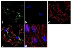
- Immunofluorescence analysis of Raptor was performed using 70% confluent log phase HeLa cells. The cells were fixed with 4% paraformaldehyde for 10 minutes, permeabilized with 0.1% Triton™ X-100 for 10 minutes, and blocked with 2% BSA for 1 hour at room temperature. The cells were labeled with Raptor Rabbit Polyclonal Antibody (Product # 42-4000) at 2 µg/mL in 0.1% BSA and incubated for 3 hours at room temperature and then labeled with Goat anti-Rabbit IgG (H+L) Superclonal™ Secondary Antibody, Alexa Fluor® 488 conjugate (Product # A27034) a dilution of 1:2000 for 45 minutes at room temperature (Panel a: green). Nuclei (Panel b: blue) were stained with SlowFade® Gold Antifade Mountant with DAPI (Product # S36938). F-actin (Panel c: red) was stained with Alexa Fluor® 555 Rhodamine Phalloidin (Product # R415, 1:300). Panel d represents the merged image showing punctate cytoplasmic localization. Panel e shows the no primary antibody control. The images were captured at 60X magnification.
- Submitted by
- Invitrogen Antibodies (provider)
- Main image

- Experimental details
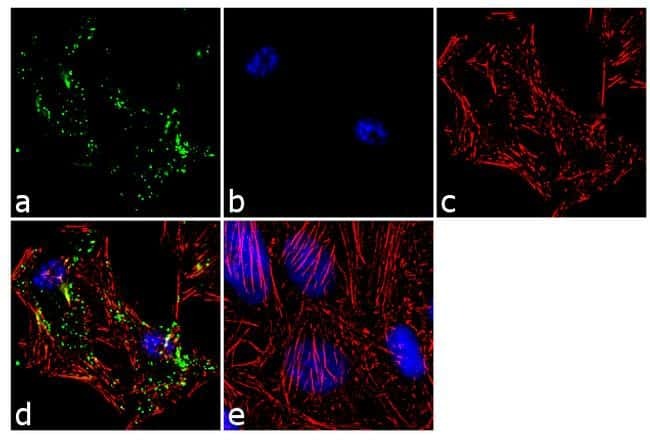
- Immunofluorescence analysis of Raptor was performed using 70% confluent log phase HeLa cells. The cells were fixed with 4% paraformaldehyde for 10 minutes, permeabilized with 0.1% Triton™ X-100 for 10 minutes, and blocked with 2% BSA for 1 hour at room temperature. The cells were labeled with Raptor Rabbit Polyclonal Antibody (Product # 42-4000) at 2 µg/mL in 0.1% BSA and incubated for 3 hours at room temperature and then labeled with Goat anti-Rabbit IgG (Heavy Chain) Superclonal™ Secondary Antibody, Alexa Fluor® 488 conjugate (Product # A27034) a dilution of 1:2000 for 45 minutes at room temperature (Panel a: green). Nuclei (Panel b: blue) were stained with SlowFade® Gold Antifade Mountant with DAPI (Product # S36938). F-actin (Panel c: red) was stained with Alexa Fluor® 555 Rhodamine Phalloidin (Product # R415, 1:300). Panel d represents the merged image showing punctate cytoplasmic localization. Panel e shows the no primary antibody control. The images were captured at 60X magnification.

Supportive validation
- Submitted by
- Invitrogen Antibodies (provider)
- Main image

- Experimental details
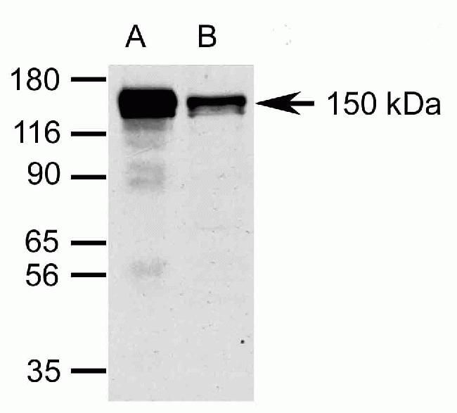
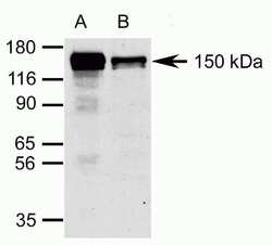
- Immunoprecipitation (A) and Western blot following immunoprecipitation (B) analysis of 293 cell lysates using Zymed (R) Rb anti-Raptor (Product # 42-4000). Image courtesy of Dr. Jeremy Copp, The Salk Institute, San Diego, CA.
Supportive validation
- Submitted by
- Invitrogen Antibodies (provider)
- Main image

- Experimental details
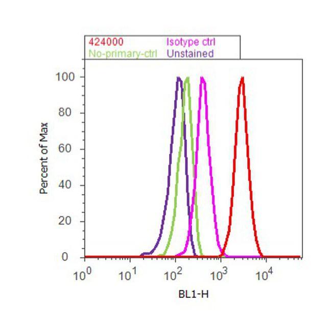
- Flow cytometry analysis of Raptor was done on HeLa cells. Cells were fixed with 70% ethanol for 10 minutes, permeabilized with 0.25% Triton™ X-100 for 20 minutes, and blocked with 5% BSA for 30 minutes at room temperature. Cells were labeled with Raptor Rabbit Polyclonal Antibody (424000, red histogram) or with rabbit isotype control (pink histogram) at 3-5 ug/million cells in 2.5% BSA. After incubation at room temperature for 2 hours, the cells were labeled with Alexa Fluor® 488 Goat Anti-Rabbit Secondary Antibody (A11008) at a dilution of 1:400 for 30 minutes at room temperature. The representative 10,000 cells were acquired and analyzed for each sample using an Attune® Acoustic Focusing Cytometer. The purple histogram represents unstained control cells and the green histogram represents no-primary-antibody control.
Supportive validation
- Submitted by
- Invitrogen Antibodies (provider)
- Main image
- Experimental details
- Immunoprecipitation (A) and Western blot following immunoprecipitation (B) analysis of 293 cell lysates using Zymed (R) Rb anti-Raptor (Product # 42-4000). Image courtesy of Dr. Jeremy Copp, The Salk Institute, San Diego, CA.

- Submitted by
- Invitrogen Antibodies (provider)
- Main image
- Experimental details
- NULL
- Submitted by
- Invitrogen Antibodies (provider)
- Main image
- Experimental details
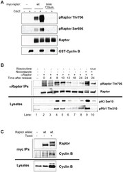
- Figure 4 Cdc2 is the raptor Ser696, Thr706 kinase. (A) Purified cdc2 can directly phosphorylate raptor on Thr706 and Ser696 in vitro. Myc-raptor (wild-type or S696/T706AA) was immunoprecipitated from hydroxyurea treated HEK293T cells. Immunoprecipitates were incubated with or without active recombinant Cdc2/cyclin B and immunoblotted with phospho-raptor Ser696, Thr706 or total raptor. (B) Endogenous raptor is phosphorylated on Thr706 in synchronized cells undergoing mitosis, and this phosphorylation is blocked by the CDK inhibitor roscovitine. A549 cells were synchronized by double thymidine block and endogenous raptor was immunoprecipitated at the indicated times after release with an anti-raptor antibody and immunoblotted with phospho-raptor Thr706. Whole cell lysates taken from the same cells were immunoblotted for mitotic markers phospho-histone H3 Ser10 and phospho-Plk1 Thr210. (C) Raptor immunoprecipitates with endogenous cyclin B. Hela cells stably expressing myc-wt raptor with stable knockdown of endogenous raptor treated with or without taxol for 16 hours. myc-tagged raptor was immunoprecipitated and immunoblotted for Cyclin B.
- Submitted by
- Invitrogen Antibodies (provider)
- Main image
- Experimental details
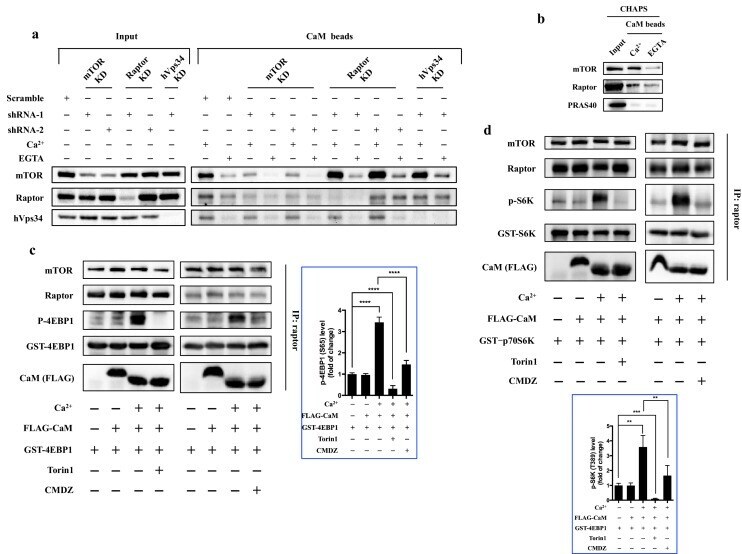
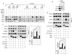
- Figure 4. CaM interacts with mTORC1 and regulates mTORC1 kinase activity in vitro. ( a ) CaM interacts with mTOR independent of hVps34 or raptor. Cell lysates were prepared from HEK293T cells transduced with lentiviral shRNAs targeting human mTOR, raptor, hVps34 or scrambled shRNA, followed by CaM sepharose precipitation in the presence of CaCl 2 (1 mM) or EGTA (5 mM). The cell lysates and precipitates were analyzed by immunoblotting to detect the indicated proteins. ( b ) Endogenous mTORC1 was pulled down by CaM sepharose in a Ca 2+ -dependent manner. HEK293T cells were lysed in CHAPS buffer, and the lysates were incubated with CaM sepharose in the presence of CaCl 2 (1 mM) or EGTA (5 mM). The precipitates were analyzed by immunoblotting. ( c and d ) Cell lysates were prepared from HEK293T cells in CHAPS buffer, and endogenous mTORC1 was immunoprecipitated by a raptor antibody. ATP (250 muM), Torin1 (100 nM), CMDZ (8 muM), CaM (2 muM) or/and CaCl 2 (1 mM) were added into the kinase reaction as indicated. Phosphorylation of 4EBP1 ( c ) and S6K ( d ) were detected by immunoblotting. The plots show the fold of change of phosphorylation of 4EBP1 ( c ) or S6K ( d ) compared with control group (first lane) normalized by total GST-tagged protein control. (mean +- s.d., n = 6 and 5 independent experiments, respectively). DOI: http://dx.doi.org/ Figure 4--figure supplement 1. Effects of calmidazolium (CMDZ) on hVps34 depleted cells. ( a ) HEK 293T cells were transduced with lentiviru