Explore
 Validate
 Learn
Learn Western blot
 Immunoprecipitation
Antibody data
- Antibody Data
- Antigen structure
- References [0]
- Comments [0]
- Validations
- Western blot [3]
- ELISA [1]
- Immunocytochemistry [1]
- Immunohistochemistry [2]
Submit
Validation data
Reference
Comment
Report error
- Product number
- 600-401-692 - Provider product page

- Provider
- Invitrogen Antibodies
- Product name
- ROBO-1 Polyclonal Antibody
- Antibody type
- Polyclonal
- Antigen
- Synthetic peptide
- Reactivity
- Human, Mouse, Rat, Canine
- Host
- Rabbit
- Isotype
- IgG
- Vial size
- 100 µg
- Concentration
- 1 mg/mL
- Storage
- -20° C, Avoid Freeze/Thaw Cycles
No comments: Submit comment
Supportive validation
- Submitted by
- Invitrogen Antibodies (provider)
- Main image
- Experimental details
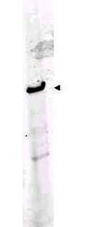
- Western blot using Rocklands Affinity Purified anti-ROBO-1 antibody shows detection of a band at ~181 kDa corresponding to ROBO-1 present in mouse brain lysate (arrowhead). Approximately 35 µg of lysate was separated by 4-8% SDS-PAGE and transferred onto nitrocellulose. After blocking the membrane was probed with the primary antibody diluted to 1:1,000. Reaction occurred 2h at room temperature followed by washes and reaction with a 1:10,000 dilution of IRDye™800 conjugated Gt-a-Rabbit IgG [H&L] MX (611-132-122) for 45 min at room temperature. IRDye™800 fluorescence image was captured using the Odyssey® Infrared Imaging System developed by LI-COR. IRDye is a trademark of LI-COR, Inc. Other detection systems will yield similar results.
- Submitted by
- Invitrogen Antibodies (provider)
- Main image
- Experimental details
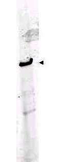
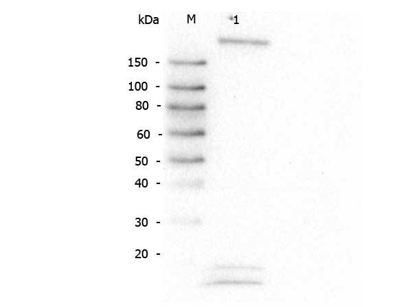
- Western Blot of Rabbit anti-Robo-1 antibody. Lane M: Super Signal Molecular Weight Marker. Lane 1: HeLa WCL. Load: 30 µg lysate. Primary antibody: Robo-1 antibody at 1:1,000 for overnight at 4°C. Secondary antibody: Peroxidase rabbit secondary antibody (p/n 611-103-122) at 1:40,000 for 30 min at RT. Block: Blocking Buffer for Fluorescent Western Blotting (p/n MB-070) for 30 min at RT. Predicted/Observed size: 181 kDa, 181 kDa for Robo-1. Other band(s): lower bands not identified.
- Submitted by
- Invitrogen Antibodies (provider)
- Main image
- Experimental details
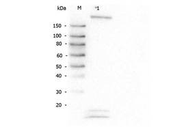
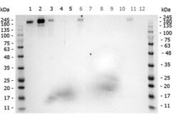
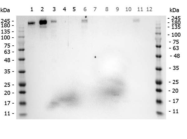
- Western Blot of Rabbit anti-ROBO1 antibody. Marker: Opal Pre-stained ladder (p/n MB-210-0500). Lane 1: HEK293 lysate (p/n W09-000-365). Lane 2: HeLa Lysate (p/n W09-000-363). Lane 3: MCF-7 Lysate (p/n W09-000-360). Lane 4: Jurkat Lysate (p/n W09-000-370). Lane 5: A431 Lysate (p/n W09-000-361). Lane 6: A549 Lysate (p/n W09-001-372). Lane 7: LNCap Lysate (p/n W09-001-GJ9). Lane 8: MOLT-4 Lysate (p/n W09-001-GK2). Lane 9: Ramos Lysate (p/n W09-000-GK4). Lane 10: Raji Lysate (p/n W09-001-368). Lane 11: A-172 Lysate (p/n W09-001-GL5). Lane 12: NIH/3T3 Lysate (p/n W10-000-358). Load: 35 µg per lane. Primary antibody: ROBO1 antibody at 1ug/mL overnight at 4C. Secondary antibody: Peroxidase rabbit secondary antibody (p/n 611-103-122) at 1:30,000 for 60 min at RT. Blocking Buffer: 1% Casein-TTBS for 30 min at RT. Predicted/Observed size: 181kDa for ROBO1.
Supportive validation
- Submitted by
- Invitrogen Antibodies (provider)
- Main image
- Experimental details
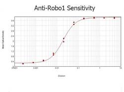
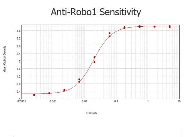
- ELISA results of purified Rabbit anti-Robo-1 Antibody tested against BSA-conjugated peptide of immunizing peptide. Each well was coated in duplicate with 0.1 µg of conjugate. The starting dilution of antibody was 5 µg/mL and the X-axis represents the Log10 of a 3-fold dilution. This titration is a 4-parameter curve fit where the IC50 is defined as the titer of the antibody. Assay performed using 3% fish gel, Goat anti-Rabbit IgG Antibody Peroxidase Conjugated (Min X Bv Ch Gt GP Ham Hs Hu Ms Rt & Sh Serum Proteins) (p/n 611-103-122) and TMB ELISA Peroxidase Substrate (p/n TMBE-1000).
Supportive validation
- Submitted by
- Invitrogen Antibodies (provider)
- Main image
- Experimental details
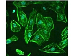
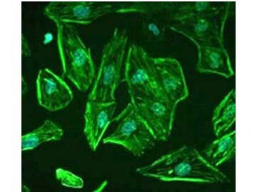
- Staining of ROBO1 in undifferentiated, immortalized human podocytes by Immunocytochemistry/ Immunofluorescence. Cells were fixed with 2% paraformaldehyde and 4% sucrose at room temperature for 10 minutes. The cells were then washed once with PBS, permeabilized with 0.3% Triton X-100 for 10 minutes and incubated with blocking solution (2% FCS, 2% BSA, 0.2% fish gelatin) for 30 minutes, before further incubation with primary Ab for 1 hour. An Alexa Fluor 488 goat anti-rabbit IgG secondary antibody was used at a dilution of 1/200. DAPI was used for nuclear counterstaining. Image from Lindenmeyer MT et al. Systematic Analysis of a Novel Human Renal Glomerulus-Enriched Gene Expression Dataset. PLoS One. 2010 July 12;5(7):e11545, Fig 5.
Supportive validation
- Submitted by
- Invitrogen Antibodies (provider)
- Main image
- Experimental details
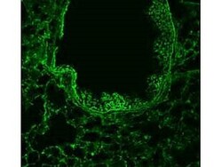
- Immunofluorescence of Anti-ROBO-1 Antibody. 1/50 staining mouse lung tissue sections (adult, frozen 100µm wholemount sections) by IHC-Fr. The tissue was paraformaldehyde fixed and permeabilized with triton x-100 before incubation with the antibody for 16 hours at 4°C.

- Submitted by
- Invitrogen Antibodies (provider)
- Main image

- Experimental details
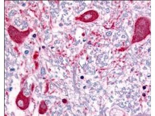
- Rocklands Affinity Purified anti-ROBO1 antibody was used at a concentration of 5 µg/ml to detect ROBO1 in a variety of tissues including multi-human, multi-brain and multi-cancer slides. This image shows staining of human brain tissue. Tissue was formalin-fixed and paraffin embedded. Personal Communication, Tina Roush, LifeSpanBiosciences, Seattle, WA.