Explore
 Validate
 Learn
Learn Western blot
 Immunocytochemistry
Antibody data
- Antibody Data
- Antigen structure
- References [3]
- Comments [0]
- Validations
- Immunocytochemistry [4]
- Immunohistochemistry [1]
Submit
Validation data
Reference
Comment
Report error
- Product number
- 35-2900 - Provider product page

- Provider
- Invitrogen Antibodies
- Product name
- EphB4 Monoclonal Antibody (3D7F8)
- Antibody type
- Monoclonal
- Antigen
- Recombinant full-length protein
- Description
- 37-1800 has been successfully used in Western Blot, immunohistochemical, immunofluorescence, and ELISA assays. 35-2900 by western blot detects a ~125kDa band representing EphB4 but also detects a strong band at ~90kDa , likely representing another protein in the ephrin family. 37-1800 is recommended for WB.
- Reactivity
- Human
- Host
- Mouse
- Isotype
- IgG
- Antibody clone number
- 3D7F8
- Vial size
- 100 μg
- Concentration
- 0.5 mg/mL
- Storage
- -20°C
Submitted references Class I and III HDACs and loss of active chromatin features contribute to epigenetic silencing of CDX1 and EPHB tumor suppressor genes in colorectal cancer.
Class I and III HDACs and loss of active chromatin features contribute to epigenetic silencing of CDX1 and EPHB tumor suppressor genes in colorectal cancer.
The EphA4 receptor regulates neuronal morphology through SPAR-mediated inactivation of Rap GTPases.
Rönsch K, Jäger M, Schöpflin A, Danciu M, Lassmann S, Hecht A
Epigenetics 2011 May;6(5):610-22
Epigenetics 2011 May;6(5):610-22
Class I and III HDACs and loss of active chromatin features contribute to epigenetic silencing of CDX1 and EPHB tumor suppressor genes in colorectal cancer.
Rönsch K, Jäger M, Schöpflin A, Danciu M, Lassmann S, Hecht A
Epigenetics 2011 May;6(5):610-22
Epigenetics 2011 May;6(5):610-22
The EphA4 receptor regulates neuronal morphology through SPAR-mediated inactivation of Rap GTPases.
Richter M, Murai KK, Bourgin C, Pak DT, Pasquale EB
The Journal of neuroscience : the official journal of the Society for Neuroscience 2007 Dec 19;27(51):14205-15
The Journal of neuroscience : the official journal of the Society for Neuroscience 2007 Dec 19;27(51):14205-15
No comments: Submit comment
Supportive validation
- Submitted by
- Invitrogen Antibodies (provider)
- Main image

- Experimental details
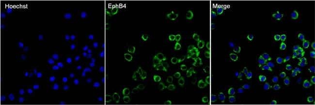
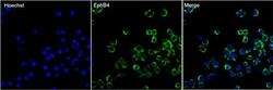
- Immunofluorescent analysis of EphB4 (green) in HT29 cells. The cells were fixed with 4% paraformaldehyde in PBS for 15 minutes at room temperature and blocked with 3% BSA for 30 minutes at room temperature. Cells were stained with a EphB4 mouse monoclonal antibody (Product # 35-2900) at a concentration of 5 µg/mL in blocking buffer for 1 hour at room temperature, and then incubated with a Goat anti-Mouse IgG (H+L) Secondary Antibody, Alexa Fluor Plus 488 conjugate (Product # A32731) at a dilution of 1:500 for at least 30 minutes at a room temperature in the dark (green). Nuclei (blue) were stained with Hoechst 33342 (Product # 62249). Images were taken on a Thermo Scientific ToxInsight Instrument at 20X magnification.
- Submitted by
- Invitrogen Antibodies (provider)
- Main image

- Experimental details
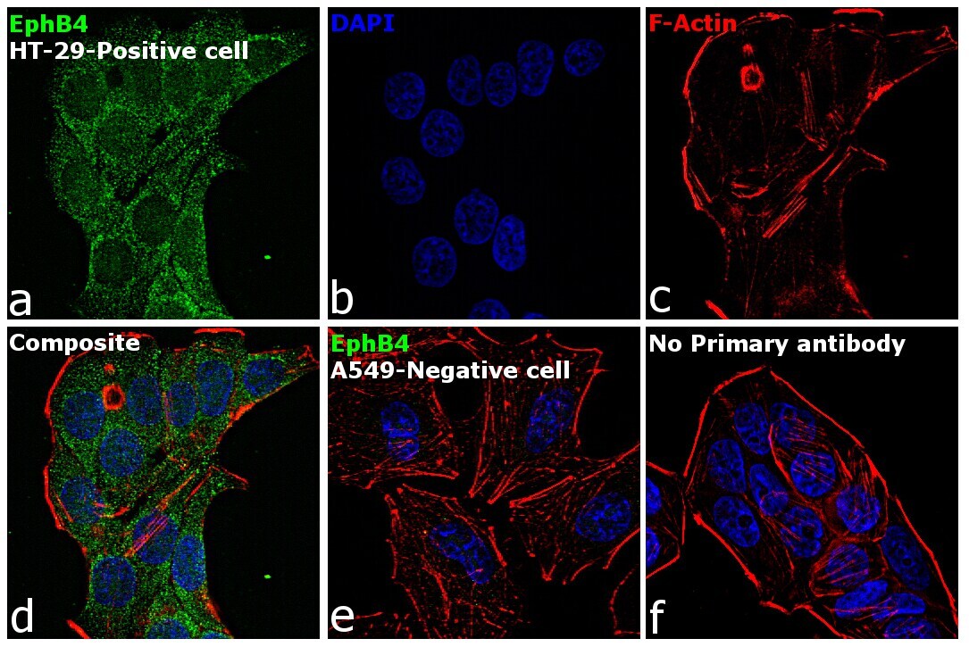
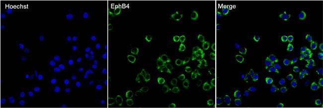
- Immunofluorescence analysis of EphB4 was performed using 70% confluent log phase HT-29, A549 cells. The cells were fixed with 4% paraformaldehyde for 10 minutes, permeabilized with 0.1% Triton™ X-100 for 15 minutes, and blocked with 2% BSA for 1 hour at room temperature. The cells were labeled with EphB4 Mouse Monoclonal Antibody (3D7F8) (Product # 35-2900) at 4 µg/mL in 0.1% BSA, incubated at 4 degree celsius overnight and then with Goat anti-Mouse IgG (H+L) Superclonal™ Recombinant Secondary Antibody, Alexa Fluor® 488 conjugate (Product # A28175) at a dilution of 1:2000 for 45 minutes at room temperature (Panel a: green). Nuclei (Panel b: blue) were stained with ProLong™ Diamond Antifade Mountant with DAPI (Product # P36962). F-actin (Panel c: red) was stained with Rhodamine Phalloidin (Product # R415, 1:300). Panel d represents the merged image showing Cytoplasmic localization. Panel e represents A549 cells having no expression of EphB4. Panel f represents control cells with no primary antibody to assess background. The images were captured at 60X magnification.
- Submitted by
- Invitrogen Antibodies (provider)
- Main image
- Experimental details
- Immunofluorescence analysis of EphB4 was performed using 70% confluent log phase HT-29, A549 cells. The cells were fixed with 4% paraformaldehyde for 10 minutes, permeabilized with 0.1% Triton™ X-100 for 15 minutes, and blocked with 2% BSA for 1 hour at room temperature. The cells were labeled with EphB4 Mouse Monoclonal Antibody (3D7F8) (Product # 35-2900) at 4 µg/mL in 0.1% BSA, incubated at 4 degree celsius overnight and then with Goat anti-Mouse IgG (H+L) Superclonal™ Recombinant Secondary Antibody, Alexa Fluor® 488 conjugate (Product # A28175) at a dilution of 1:2000 for 45 minutes at room temperature (Panel a: green). Nuclei (Panel b: blue) were stained with ProLong™ Diamond Antifade Mountant with DAPI (Product # P36962). F-actin (Panel c: red) was stained with Rhodamine Phalloidin (Product # R415, 1:300). Panel d represents the merged image showing Cytoplasmic localization. Panel e represents A549 cells having no expression of EphB4. Panel f represents control cells with no primary antibody to assess background. The images were captured at 60X magnification.

- Submitted by
- Invitrogen Antibodies (provider)
- Main image
- Experimental details
- Immunofluorescent analysis of EphB4 (green) in HT29 cells. The cells were fixed with 4% paraformaldehyde in PBS for 15 minutes at room temperature and blocked with 3% BSA for 30 minutes at room temperature. Cells were stained with a EphB4 mouse monoclonal antibody (Product # 35-2900) at a concentration of 5 µg/mL in blocking buffer for 1 hour at room temperature, and then incubated with a Goat anti-Mouse IgG (H+L) Secondary Antibody, Alexa Fluor Plus 488 conjugate (Product # A32731) at a dilution of 1:500 for at least 30 minutes at a room temperature in the dark (green). Nuclei (blue) were stained with Hoechst 33342 (Product # 62249). Images were taken on a Thermo Scientific ToxInsight Instrument at 20X magnification.
Supportive validation
- Submitted by
- Invitrogen Antibodies (provider)
- Main image
- Experimental details
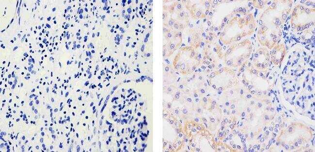
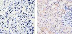
- Immunohistochemistry analysis of EPHB4 RECEPTOR showing staining in the cytoplasm of paraffin-embedded human kidney tissue (right) compared to a negative control without primary antibody (left). To expose target proteins, antigen retrieval was performed using 10mM sodium citrate (pH 6.0), microwaved for 8-15 min. Following antigen retrieval, tissues were blocked in 3% H2O2-methanol for 15 min at room temperature, washed with ddH2O and PBS, and then probed with a EPHB4 RECEPTOR Mouse Monoclonal Antibody (Product # 35-2900) diluted in 3% BSA-PBS at a dilution of 1:20 for 1 hour at 37ºC in a humidified chamber. Tissues were washed extensively in PBST and detection was performed using an HRP-conjugated secondary antibody followed by colorimetric detection using a DAB kit. Tissues were counterstained with hematoxylin and dehydrated with ethanol and xylene to prep for mounting.