Explore
 Validate
 Learn
Learn Western blot
Antibody data
- Antibody Data
- Antigen structure
- References [6]
- Comments [0]
- Validations
- Western blot [3]
- Immunohistochemistry [1]
- Flow cytometry [1]
Submit
Validation data
Reference
Comment
Report error
- Product number
- AF3038 - Provider product page

- Provider
- R&D Systems
- Product name
- Human EphB4 Antibody
- Antibody type
- Polyclonal
- Description
- Antigen Affinity-purified. Detects human EphB4 in direct ELISAs and Western blots. In direct ELISAs, less than 10% cross-reactivity with recombinant mouse (rm) EphB4 is observed and less than 1% cross-reactivity with recombinant human (rh) EphA4, rhEphA3, rhEphB2, and rhEphB3 is observed.
- Reactivity
- Human
- Host
- Goat
- Conjugate
- Unconjugated
- Antigen sequence
P54760- Isotype
- IgG
- Vial size
- 100 ug
- Concentration
- LYOPH
- Storage
- Use a manual defrost freezer and avoid repeated freeze-thaw cycles. 12 months from date of receipt, -20 to -70 °C as supplied. 1 month, 2 to 8 °C under sterile conditions after reconstitution. 6 months, -20 to -70 °C under sterile conditions after reconstitution.
Submitted references Downregulation of proinflammatory cytokines in HTLV-1-infected T cells by Resveratrol.
EPHB4 kinase-inactivating mutations cause autosomal dominant lymphatic-related hydrops fetalis.
Instruction of circulating endothelial progenitors in vitro towards specialized blood-brain barrier and arterial phenotypes.
BCR-ABL-independent and RAS / MAPK pathway-dependent form of imatinib resistance in Ph-positive acute lymphoblastic leukemia cell line with activation of EphB4.
Soluble EphB4 inhibition of PDGF-induced RPE migration in vitro.
Activation of the receptor EphB4 by its specific ligand ephrin B2 in human osteoarthritic subchondral bone osteoblasts.
Fuggetta MP, Bordignon V, Cottarelli A, Macchi B, Frezza C, Cordiali-Fei P, Ensoli F, Ciafrè S, Marino-Merlo F, Mastino A, Ravagnan G
Journal of experimental & clinical cancer research : CR 2016 Jul 22;35(1):118
Journal of experimental & clinical cancer research : CR 2016 Jul 22;35(1):118
EPHB4 kinase-inactivating mutations cause autosomal dominant lymphatic-related hydrops fetalis.
Martin-Almedina S, Martinez-Corral I, Holdhus R, Vicente A, Fotiou E, Lin S, Petersen K, Simpson MA, Hoischen A, Gilissen C, Jeffery H, Atton G, Karapouliou C, Brice G, Gordon K, Wiseman JW, Wedin M, Rockson SG, Jeffery S, Mortimer PS, Snyder MP, Berland S, Mansour S, Makinen T, Ostergaard P
The Journal of clinical investigation 2016 Aug 1;126(8):3080-8
The Journal of clinical investigation 2016 Aug 1;126(8):3080-8
Instruction of circulating endothelial progenitors in vitro towards specialized blood-brain barrier and arterial phenotypes.
Boyer-Di Ponio J, El-Ayoubi F, Glacial F, Ganeshamoorthy K, Driancourt C, Godet M, Perrière N, Guillevic O, Couraud PO, Uzan G
PloS one 2014;9(1):e84179
PloS one 2014;9(1):e84179
BCR-ABL-independent and RAS / MAPK pathway-dependent form of imatinib resistance in Ph-positive acute lymphoblastic leukemia cell line with activation of EphB4.
Suzuki M, Abe A, Imagama S, Nomura Y, Tanizaki R, Minami Y, Hayakawa F, Ito Y, Katsumi A, Yamamoto K, Emi N, Kiyoi H, Naoe T
European journal of haematology 2010 Mar;84(3):229-38
European journal of haematology 2010 Mar;84(3):229-38
Soluble EphB4 inhibition of PDGF-induced RPE migration in vitro.
He S, Kumar SR, Zhou P, Krasnoperov V, Ryan SJ, Gill PS, Hinton DR
Investigative ophthalmology & visual science 2010 Jan;51(1):543-52
Investigative ophthalmology & visual science 2010 Jan;51(1):543-52
Activation of the receptor EphB4 by its specific ligand ephrin B2 in human osteoarthritic subchondral bone osteoblasts.
Kwan Tat S, Pelletier JP, Amiable N, Boileau C, Lajeunesse D, Duval N, Martel-Pelletier J
Arthritis and rheumatism 2008 Dec;58(12):3820-30
Arthritis and rheumatism 2008 Dec;58(12):3820-30
No comments: Submit comment
Supportive validation
- Submitted by
- R&D Systems (provider)
- Main image
- Experimental details
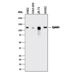
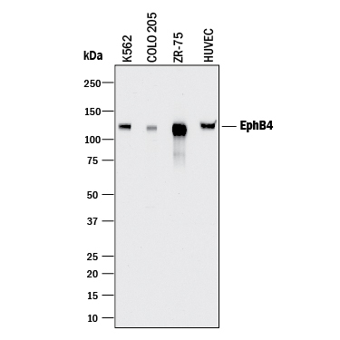
- Detection of Human EphB4 by Western Blot. Western blot shows lysates of K562 human chronic myelogenous leukemia cell line, COLO 205 human colorectal adenocarcinoma cell line, ZR-75 human breast cancer cell line, and HUVEC human umbilical vein endothelial cells. PVDF membrane was probed with 2 µg/mL of Goat Anti-Human EphB4 Antigen Affinity-purified Polyclonal Antibody (Catalog # AF3038) followed by HRP-conjugated Anti-Goat IgG Secondary Antibody (Catalog # HAF017). A specific band was detected for EphB4 at approximately 120 kDa (as indicated). This experiment was conducted under reducing conditions and using Immunoblot Buffer Group 1.
- Submitted by
- R&D Systems (provider)
- Main image
- Experimental details
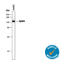
- Detection of Human EphB4 by Simple WesternTM. Simple Western lane view shows lysates of HUVEC human umbilical vein endothelial cells, loaded at 0.2 mg/mL. A specific band was detected for EphB4 at approximately 127 kDa (as indicated) using 50 µg/mL of Goat Anti-Human EphB4 Antigen Affinity-purified Polyclonal Antibody (Catalog # AF3038) followed by 1:50 dilution of HRP-conjugated Anti-Goat IgG Secondary Antibody (Catalog # HAF109). This experiment was conducted under reducing conditions and using the 12-230 kDa separation system.

- Submitted by
- R&D Systems (provider)
- Main image
- Experimental details
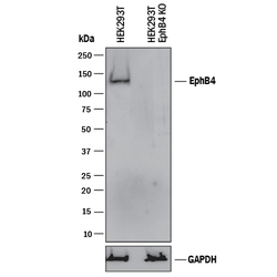
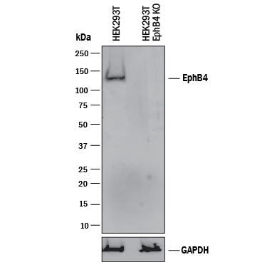
- Western Blot Shows Human EphB4 Specificity by Using Knockout Cell Line. Western blot shows lysates of HEK293T human embryonic kidney parental cell line and EphB4 knockout HEK293T cell line (KO). PVDF membrane was probed with 2 µg/mL of Goat Anti-Human EphB4 Antigen Affinity-purified Polyclonal Antibody (Catalog # AF3038) followed by HRP-conjugated Anti-Goat IgG Secondary Antibody (Catalog # HAF017). A specific band was detected for EphB4 at approximately 140 kDa (as indicated) in the parental HEK293T cell line, but is not detectable in knockout HEK293T cell line. GAPDH (Catalog # AF5718) is shown as a loading control. This experiment was conducted under reducing conditions and using Immunoblot Buffer Group 1.
Supportive validation
- Submitted by
- R&D Systems (provider)
- Main image
- Experimental details
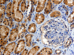
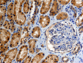
- EphB4 in Human Kidney. EphB4 was detected in immersion fixed paraffin-embedded sections of human kidney using 15 µg/mL Goat Anti-Human EphB4 Antigen Affinity-purified Polyclonal Antibody (Catalog # AF3038) overnight at 4 °C. Tissue was stained with the Anti-Goat HRP-DAB Cell & Tissue Staining Kit (brown; Catalog # CTS008) and counterstained with hematoxylin (blue). View our protocol for Chromogenic IHC Staining of Paraffin-embedded Tissue Sections.
Supportive validation
- Submitted by
- R&D Systems (provider)
- Main image
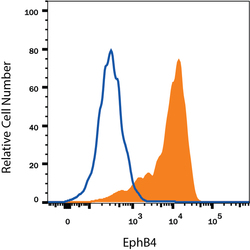
- Experimental details
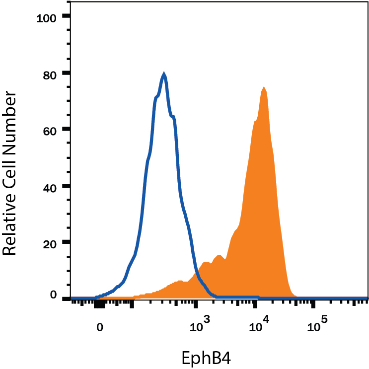
- Detection of EphB4 in MCF-7 Human Cell Line by Flow Cytometry. MCF-7 human breast cancer cell line was stained with Goat Anti-Human EphB4 Antigen Affinity-purified Polyclonal Antibody (Catalog # AF3038, filled histogram) or isotype control antibody (Catalog # AB-108-C, open histogram), followed by Phycoerythrin-conjugated Anti-Goat IgG Secondary Antibody (Catalog # F0107). View our protocol for Staining Membrane-associated Proteins.