Explore
 Validate
 Learn
Learn20639-1-AP
antibody from Proteintech Group
Targeting: CHCHD6
CHCM1, MGC13016, Mic25, MICOS25, PPP1R23
 Western blot
 ELISA
ELISAAntibody data
- Antibody Data
- Antigen structure
- References [17]
- Comments [0]
- Validations
- Western blot [1]
- Immunohistochemistry [2]
Submit
Validation data
Reference
Comment
Report error
- Product number
- 20639-1-AP - Provider product page

- Provider
- Proteintech Group
- Proper citation
- Proteintech Cat#20639-1-AP, RRID:AB_10697667
- Product name
- CHCHD6 antibody
- Antibody type
- Polyclonal
- Description
- KD/KO validated CHCHD6 antibody (Cat. #20639-1-AP) is a rabbit polyclonal antibody that shows reactivity with human and has been validated for the following applications: IHC, IP, WB, ELISA.
- Reactivity
- Human
- Host
- Rabbit
- Conjugate
- Unconjugated
- Isotype
- IgG
- Vial size
- 20ul, 150ul
Submitted references Mitochondria serve as a source of mineral precursors initiating early cartilage calcification in osteoarthritis.
Protein domain characterization reveals human MIC60 tolerates loss of helical bundle domain.
H(2)S remodels mitochondrial ultrastructure and destabilizes respiratory supercomplexes.
BRD4-mediated epigenetic regulation of endoplasmic reticulum-mitochondria contact sites is governed by the mitochondrial complex III.
H (2) S remodels mitochondrial ultrastructure and destabilizes respiratory supercomplexes.
Mitochondrial apolipoprotein MIC26 is a metabolic rheostat regulating central cellular fuel pathways.
SLP2 and MIC13 synergistically coordinate MICOS assembly and crista junction formation.
The role of the mitochondrial outer membrane protein SLC25A46 in mitochondrial fission and fusion.
The outer mitochondrial membrane protein TMEM11 demarcates spatially restricted BNIP3/BNIP3L-mediated mitophagy.
A CHCHD6-APP axis connects amyloid and mitochondrial pathology in Alzheimer's disease.
Conserved GxxxG and WN motifs of MIC13 are essential for bridging two MICOS subcomplexes.
MIC26 and MIC27 cooperate to regulate cardiolipin levels and the landscape of OXPHOS complexes.
Cristae undergo continuous cycles of membrane remodelling in a MICOS-dependent manner.
QIL1 mutation causes MICOS disassembly and early onset fatal mitochondrial encephalopathy with liver disease.
Mic60/Mitofilin determines MICOS assembly essential for mitochondrial dynamics and mtDNA nucleoid organization.
Mitofilin and CHCHD6 physically interact with Sam50 to sustain cristae structure.
QIL1 is a novel mitochondrial protein required for MICOS complex stability and cristae morphology.
Wan Q, Ye T, Gu J, Ma Y, Qin W, Yan J, Rao J, Chen L, Hao D, Tay FR, Jiao K, Niu L
Science bulletin 2026 May 15;71(9):2300-2315
Science bulletin 2026 May 15;71(9):2300-2315
Protein domain characterization reveals human MIC60 tolerates loss of helical bundle domain.
Rockfield SM, Venkataraman K, Wu CH, Wakefield R, Wu A, Budhraja A, Rodriguez-Enriquez R, Khalighifar A, Robinson CG, Li C, Carisey AF, Opferman JT
bioRxiv : the preprint server for biology 2026 Mar 22;
bioRxiv : the preprint server for biology 2026 Mar 22;
H(2)S remodels mitochondrial ultrastructure and destabilizes respiratory supercomplexes.
Hanna DA, Chen B, Shah YM, Khalimonchuk O, Cunniff B, Banerjee R
The Journal of biological chemistry 2025 May;301(5):108433
The Journal of biological chemistry 2025 May;301(5):108433
BRD4-mediated epigenetic regulation of endoplasmic reticulum-mitochondria contact sites is governed by the mitochondrial complex III.
Chen B, Stark DC, Jadhav PV, Lynn-Nguyen TM, Halligan BS, Rossiter NJ, Sindoni N, Shin M, Paulo JA, Chang M, Koo I, Koshkin S, Eyunni S, Ronchi P, Paulsen MT, Morlacchi P, Hanna DA, Lin J, Guerra RM, Pagliarini DJ, Banerjee R, Parolia A, Ljungman ME, Patterson AD, Mancias JD, Mosalaganti S, Sexton JZ, Calì T, Lyssiotis CA, Shah YM
bioRxiv : the preprint server for biology 2024 Nov 5;
bioRxiv : the preprint server for biology 2024 Nov 5;
H (2) S remodels mitochondrial ultrastructure and destabilizes respiratory supercomplexes.
Hanna DA, Chen B, Shah YM, Khalimonchuk O, Cunniff B, Banerjee R
bioRxiv : the preprint server for biology 2024 Nov 3;
bioRxiv : the preprint server for biology 2024 Nov 3;
Mitochondrial apolipoprotein MIC26 is a metabolic rheostat regulating central cellular fuel pathways.
Damiecki M, Naha R, Schaumkessel Y, Westhoff P, Atanelov N, Stefanski A, Petzsch P, Stühler K, Köhrer K, Weber AP, Anand R, Reichert AS, Kondadi AK
Life science alliance 2024 Dec;7(12)
Life science alliance 2024 Dec;7(12)
SLP2 and MIC13 synergistically coordinate MICOS assembly and crista junction formation.
Naha R, Strohm R, Schaumkessel Y, Urbach J, Wittig I, Reichert AS, Kondadi AK, Anand R
iScience 2024 Dec 20;27(12):111467
iScience 2024 Dec 20;27(12):111467
The role of the mitochondrial outer membrane protein SLC25A46 in mitochondrial fission and fusion.
Schuettpelz J, Janer A, Antonicka H, Shoubridge EA
Life science alliance 2023 Jun;6(6)
Life science alliance 2023 Jun;6(6)
The outer mitochondrial membrane protein TMEM11 demarcates spatially restricted BNIP3/BNIP3L-mediated mitophagy.
Gok MO, Connor OM, Wang X, Menezes CJ, Llamas CB, Mishra P, Friedman JR
The Journal of cell biology 2023 Apr 3;222(4)
The Journal of cell biology 2023 Apr 3;222(4)
A CHCHD6-APP axis connects amyloid and mitochondrial pathology in Alzheimer's disease.
Shang Y, Sun X, Chen X, Wang Q, Wang EJ, Miller E, Xu R, Pieper AA, Qi X
Acta neuropathologica 2022 Nov;144(5):911-938
Acta neuropathologica 2022 Nov;144(5):911-938
Conserved GxxxG and WN motifs of MIC13 are essential for bridging two MICOS subcomplexes.
Urbach J, Kondadi AK, David C, Naha R, Deinert K, Reichert AS, Anand R
Biochimica et biophysica acta. Biomembranes 2021 Dec 1;1863(12):183683
Biochimica et biophysica acta. Biomembranes 2021 Dec 1;1863(12):183683
MIC26 and MIC27 cooperate to regulate cardiolipin levels and the landscape of OXPHOS complexes.
Anand R, Kondadi AK, Meisterknecht J, Golombek M, Nortmann O, Riedel J, Peifer-Weiß L, Brocke-Ahmadinejad N, Schlütermann D, Stork B, Eichmann TO, Wittig I, Reichert AS
Life science alliance 2020 Oct;3(10)
Life science alliance 2020 Oct;3(10)
Cristae undergo continuous cycles of membrane remodelling in a MICOS-dependent manner.
Kondadi AK, Anand R, Hänsch S, Urbach J, Zobel T, Wolf DM, Segawa M, Liesa M, Shirihai OS, Weidtkamp-Peters S, Reichert AS
EMBO reports 2020 Mar 4;21(3):e49776
EMBO reports 2020 Mar 4;21(3):e49776
QIL1 mutation causes MICOS disassembly and early onset fatal mitochondrial encephalopathy with liver disease.
Guarani V, Jardel C, Chrétien D, Lombès A, Bénit P, Labasse C, Lacène E, Bourillon A, Imbard A, Benoist JF, Dorboz I, Gilleron M, Goetzman ES, Gaignard P, Slama A, Elmaleh-Bergès M, Romero NB, Rustin P, Ogier de Baulny H, Paulo JA, Harper JW, Schiff M
eLife 2016 Sep 13;5
eLife 2016 Sep 13;5
Mic60/Mitofilin determines MICOS assembly essential for mitochondrial dynamics and mtDNA nucleoid organization.
Li H, Ruan Y, Zhang K, Jian F, Hu C, Miao L, Gong L, Sun L, Zhang X, Chen S, Chen H, Liu D, Song Z
Cell death and differentiation 2016 Mar;23(3):380-92
Cell death and differentiation 2016 Mar;23(3):380-92
Mitofilin and CHCHD6 physically interact with Sam50 to sustain cristae structure.
Ding C, Wu Z, Huang L, Wang Y, Xue J, Chen S, Deng Z, Wang L, Song Z, Chen S
Scientific reports 2015 Nov 4;5:16064
Scientific reports 2015 Nov 4;5:16064
QIL1 is a novel mitochondrial protein required for MICOS complex stability and cristae morphology.
Guarani V, McNeill EM, Paulo JA, Huttlin EL, Fröhlich F, Gygi SP, Van Vactor D, Harper JW
eLife 2015 May 21;4
eLife 2015 May 21;4
No comments: Submit comment
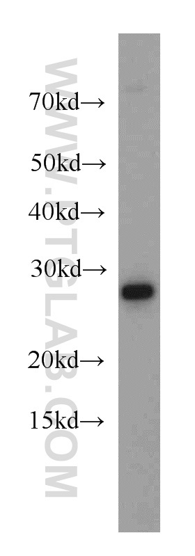
Supportive validation
- Submitted by
- Proteintech Group (provider)
- Main image

- Experimental details
- HeLa cells were subjected to SDS PAGE followed by western blot with 20639-1-AP(CHCHD6 antibody) at dilution of 1:500
- Sample type
- cell line
Supportive validation
Supportive validation
- Submitted by
- Proteintech Group (provider)
- Main image
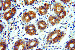
- Experimental details
- The CHCHD6 antibody from Proteintech is a rabbit polyclonal antibody to a fusion protein of human CHCHD6. This antibody recognizes human antigen. The CHCHD6 antibody has been validated for the following applications: ELISA, WB, IHC analysis.
Supportive validation
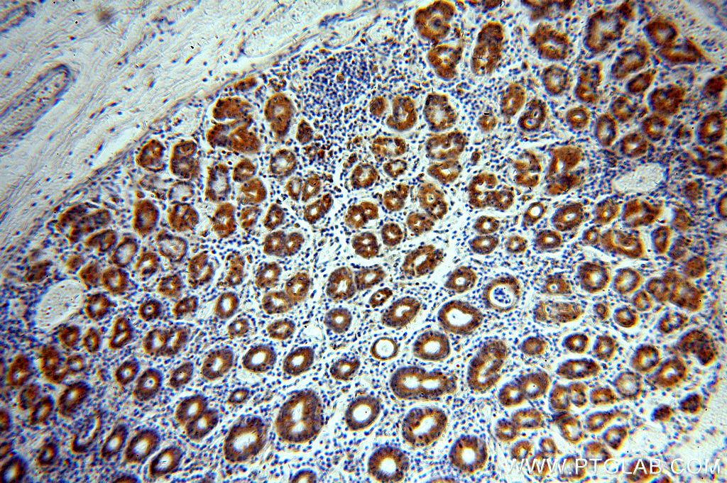
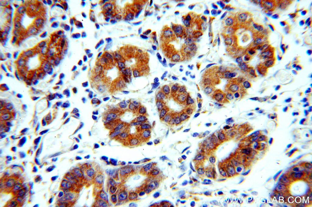
- Submitted by
- Proteintech Group (provider)
- Main image
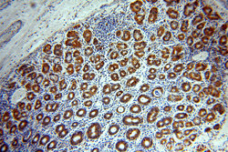
- Experimental details
- Immunohistochemical of paraffin-embedded human stomach using 20639-1-AP(CHCHD6 antibody) at dilution of 1:100 (under 10x lens)
- Sample type
- tissue