 Explore
 Validate
 Learn
Learn Immunocytochemistry
 Flow cytometry
Antibody data
- Antibody Data
- Antigen structure
- References [2]
- Comments [0]
- Validations
- Immunocytochemistry [1]
Submit
Validation data
Reference
Comment
Report error
- Product number
- MAB1524 - Provider product page

- Provider
- R&D Systems
- Product name
- Human Endoglycan/PODXL2 Antibody
- Antibody type
- Monoclonal
- Description
- Protein A or G purified from hybridoma culture supernatant. Detects human Endoglycan/PODXL2 in direct ELISAs.
- Reactivity
- Human
- Host
- Mouse
- Conjugate
- Unconjugated
- Antigen sequence
Q9NZ53- Isotype
- IgG
- Antibody clone number
- 211816
- Vial size
- 100 ug
- Concentration
- LYOPH
- Storage
- Use a manual defrost freezer and avoid repeated freeze-thaw cycles. 12 months from date of receipt, -20 to -70 °C as supplied. 1 month, 2 to 8 °C under sterile conditions after reconstitution. 6 months, -20 to -70 °C under sterile conditions after reconstitution.
Submitted references Endoglycan, a member of the CD34 family of sialomucins, is a ligand for the vascular selectins.
Endoglycan, a member of the CD34 family of sialomucins, is a ligand for the vascular selectins.
Kerr SC, Fieger CB, Snapp KR, Rosen SD
Journal of immunology (Baltimore, Md. : 1950) 2008 Jul 15;181(2):1480-90
Journal of immunology (Baltimore, Md. : 1950) 2008 Jul 15;181(2):1480-90
Endoglycan, a member of the CD34 family of sialomucins, is a ligand for the vascular selectins.
Kerr SC, Fieger CB, Snapp KR, Rosen SD
Journal of immunology (Baltimore, Md. : 1950) 2008 Jul 15;181(2):1480-90
Journal of immunology (Baltimore, Md. : 1950) 2008 Jul 15;181(2):1480-90
No comments: Submit comment
Supportive validation
- Submitted by
- R&D Systems (provider)
- Main image
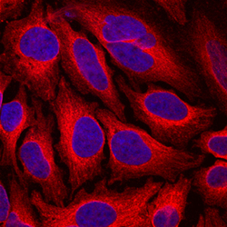
- Experimental details
- Endoglycan/PODXL2 in HeLa Human Cell Line. Endoglycan/PODXL2 was detected in immersion fixed HeLa human cervical epithelial carcinoma cell line using Mouse Anti-Human Endoglycan/PODXL2 Monoclonal Antibody (Catalog # MAB1524) at 10 µg/mL for 3 hours at room temperature. Cells were stained using the NorthernLights™ 557-conjugated Anti-Mouse IgG Secondary Antibody (red; Catalog # NL007) and counterstained with DAPI (blue). Specific staining was localized to cytoplasm. View our protocol for Fluorescent ICC Staining of Cells on Coverslips.
