Explore
 Validate
 Learn
Learn Western blot
 Immunohistochemistry
Antibody data
- Antibody Data
- Antigen structure
- References [0]
- Comments [0]
- Validations
- Western blot [1]
- Immunocytochemistry [3]
Submit
Validation data
Reference
Comment
Report error
- Product number
- MA5-45651 - Provider product page

- Provider
- Invitrogen Antibodies
- Product name
- FGF13 Monoclonal Antibody (N235/22), APC
- Antibody type
- Monoclonal
- Antigen
- Synthetic peptide
- Description
- 100% identical to rat, 94% identical to mouse. >80% identity with FGF12A/FHF1A, FGF14A/FHF4A and FGF11A/FHF3A. 1 µg/mL of MA5-45651 was sufficient for detection of FGFA/FHFA (pan) in 20 µg of rat brain lysate by colorimetric immunoblot analysis using Goat anti-mouse IgG:HRP as the secondary antibody.|Detects approximately 30kDa. Does not cross-react with FGF13B/FHF2B. Cross reacts with FGF12A/FHF1A and FGF14A/FHF4A. This antibody was formerly sold as clone S235-22.
- Reactivity
- Human, Mouse, Rat
- Host
- Mouse
- Isotype
- IgG
- Antibody clone number
- N235/22
- Vial size
- 100 μg
- Concentration
- 1 mg/mL
- Storage
- 4°C
No comments: Submit comment
Supportive validation
- Submitted by
- Invitrogen Antibodies (provider)
- Main image
- Experimental details
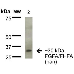
- Western Blot analysis of Rat Brain Membrane showing detection of ~30 kDa FGFA/FHFA (pan) protein. Lane 1: Molecular Weight Ladder. Lane 2: Rat Brain Membrane. Load: 15 µg. Blocking: 2% BSA and 2% Skim Milk in 1X TBST. Samples were incubated with FGFA/FHFA (pan) monoclonal antibody (Product # MA5-45651) at 1:200 for 16 hours at 4°C, followed by Goat Anti-Mouse IgG: HRP at 1:1,000 for 1 hour RT. Color Development: ECL solution for 6 min in RT. Predicted/Observed Size: ~30 kDa.
Supportive validation
- Submitted by
- Invitrogen Antibodies (provider)
- Main image
- Experimental details
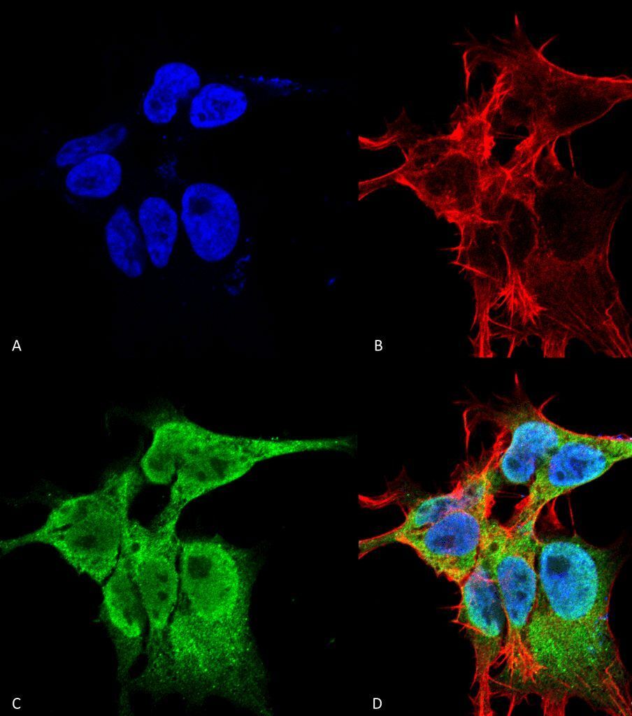
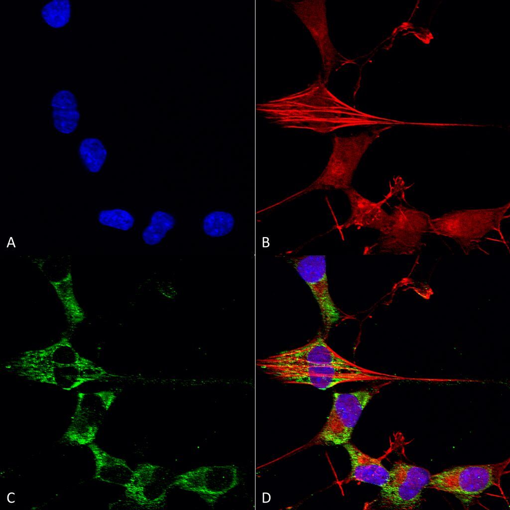
- Immunocytochemistry/Immunofluorescence analysis using human neuroblastoma cells. Fixation involved 4% PFA for 15 min. Samples were incubated with FGFA/FHFA (pan) monoclonal antibody (Product # MA5-45651) at 1:50 for overnight at 4°C with slow rocking, followed by AlexaFluor 488 at 1:1,000 for 1 hour at RT. Counterstain used was Phalloidin-iFluor 647 (red) F-Actin stain; Hoechst (blue) nuclear stain at 1:800, 1.6mM for 20 min at RT. (A) Hoechst (blue) nuclear stain. (B) Phalloidin-iFluor 647 (red) F-Actin stain. (C) FGFA/FHFA (pan) antibody (D) Composite.
- Submitted by
- Invitrogen Antibodies (provider)
- Main image
- Experimental details
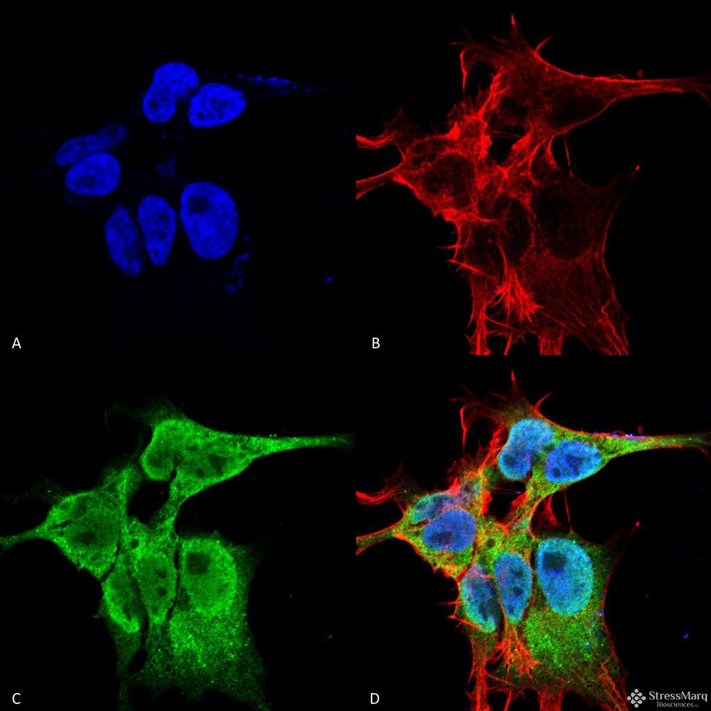
- Immunocytochemistry/Immunofluorescence analysis using human neuroblastoma cells. Fixation involved 4% Formaldehyde for 15 min at RT. Samples were incubated with FGFA/FHFA (pan) monoclonal antibody (Product # MA5-45651) at 1:100 for 60 min at RT, followed by Goat Anti-Mouse ATTO 488 at 1:100 for 60 min at RT. Counterstain used was Phalloidin Texas Red F-Actin stain; DAPI (blue) nuclear stain at 1:1,000; 1:5,000 for 60 min RT, 5 min RT. Localization: Cell Projection, Nucleus, Cytoplasm. Magnification: 60X. (A) DAPI (blue) nuclear stain. (B) Phalloidin Texas Red F-Actin stain. (C) FGFA/FHFA (pan) antibody. (D) Composite.
- Submitted by
- Invitrogen Antibodies (provider)
- Main image

- Experimental details
- Immunocytochemistry/Immunofluorescence analysis using human neuroblastoma cells. Fixation involved 4% Formaldehyde for 15 min at RT. Samples were incubated with FGFA/FHFA (pan) monoclonal antibody (Product # MA5-45651) at 1:100 for 60 min at RT, followed by Goat Anti-Mouse ATTO 488 at 1:100 for 60 min at RT. Counterstain used was Phalloidin Texas Red F-Actin stain; DAPI (blue) nuclear stain at 1:1,000; 1:5,000 for 60 min RT, 5 min RT. Localization: Cell Projection, Nucleus, Cytoplasm. Magnification: 60X. (A) DAPI (blue) nuclear stain. (B) Phalloidin Texas Red F-Actin stain. (C) FGFA/FHFA (pan) antibody. (D) Composite.