Explore
 Validate
 Learn
Learn Western blot
 Immunocytochemistry
Antibody data
- Antibody Data
- Antigen structure
- References [1]
- Comments [0]
- Validations
- Immunocytochemistry [2]
- Immunohistochemistry [4]
- Other assay [1]
Submit
Validation data
Reference
Comment
Report error
- Product number
- PA5-61244 - Provider product page

- Provider
- Invitrogen Antibodies
- Product name
- RPAP2 Polyclonal Antibody
- Antibody type
- Polyclonal
- Antigen
- Recombinant protein fragment
- Description
- Immunogen sequence: RCSRKAAGTK QTSTLKQEDA SKRKAELEAA VRKKIEFERK ALHIVEQLLE ENITEEFLME CGRFITPAHY SDVVDERSIV KLCGYPLCQK KLGI Highest antigen sequence identity to the following orthologs: Mouse - 79%, Rat - 76%.
- Reactivity
- Human
- Host
- Rabbit
- Isotype
- IgG
- Vial size
- 100 μL
- Concentration
- 0.2 mg/mL
- Storage
- Store at 4°C short term. For long term storage, store at -20°C, avoiding freeze/thaw cycles.
Submitted references Cryo-EM structure of mammalian RNA polymerase II in complex with human RPAP2.
Fianu I, Dienemann C, Aibara S, Schilbach S, Cramer P
Communications biology 2021 May 21;4(1):606
Communications biology 2021 May 21;4(1):606
No comments: Submit comment
Supportive validation
- Submitted by
- Invitrogen Antibodies (provider)
- Main image
- Experimental details
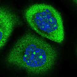
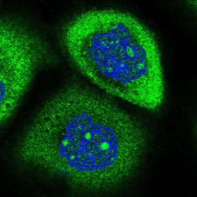
- Immunofluorescent staining of RPAP2 in human cell line A-431 shows positivity in nucleoli & cytoplasm. Samples were probed using a RPAP2 Polyclonal Antibody (Product # PA5-61244).
- Submitted by
- Invitrogen Antibodies (provider)
- Main image

- Experimental details
- Immunofluorecent analysis of RPAP2 in human cell line A-431 using RPAP2 Polyclonal Antibody (Product # PA5-61244). Staining shows positivity in nucleoli and cytoplasm.

Supportive validation
- Submitted by
- Invitrogen Antibodies (provider)
- Main image

- Experimental details
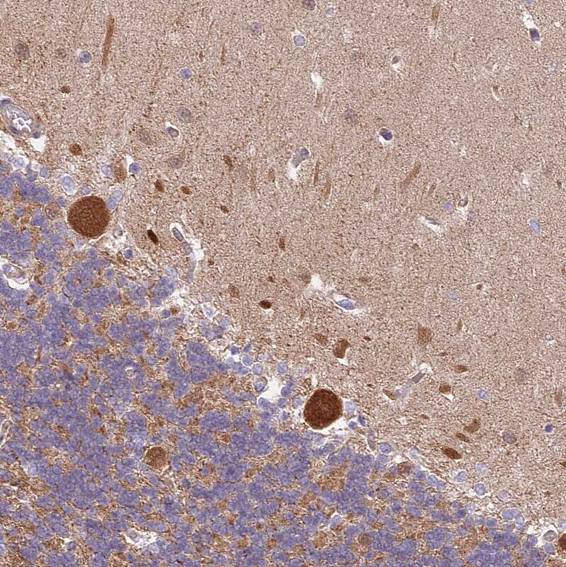
- Immunohistochemical analysis of RPAP2 in human Cerebellum using RPAP2 Polyclonal Antibody (Product # PA5-61244) shows strong nuclear and cytoplasmic positivity in Purkinje cells.
- Submitted by
- Invitrogen Antibodies (provider)
- Main image

- Experimental details
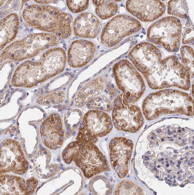
- Immunohistochemical analysis of RPAP2 in human Kidney using RPAP2 Polyclonal Antibody (Product # PA5-61244) shows moderate cytoplasmic positivity in cells in tubules.
- Submitted by
- Invitrogen Antibodies (provider)
- Main image
- Experimental details
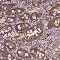
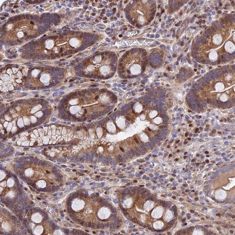
- Immunohistochemical staining of RPAP2 in human duodenum using a RPAP2 Polyclonal Antibody (Product # PA5-61244) shows moderate nuclear and cytoplasmic positivity in glandular cells.
- Submitted by
- Invitrogen Antibodies (provider)
- Main image

- Experimental details
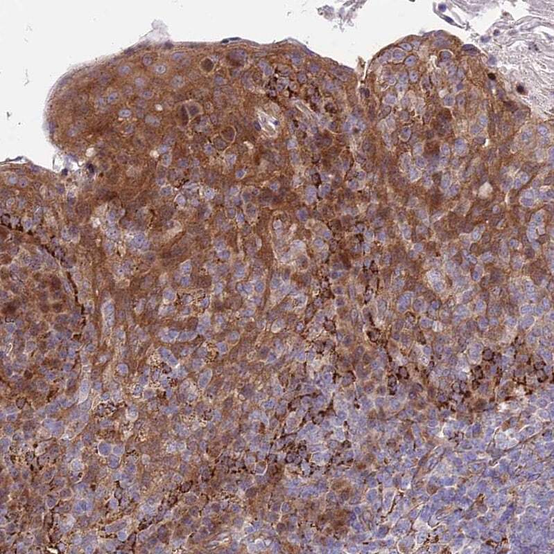
- Immunohistochemical analysis of RPAP2 in human Tonsil using RPAP2 Polyclonal Antibody (Product # PA5-61244) shows moderate nuclear and cytoplasmic positivity in squamous epithelial cells.
Supportive validation
- Submitted by
- Invitrogen Antibodies (provider)
- Main image
- Experimental details
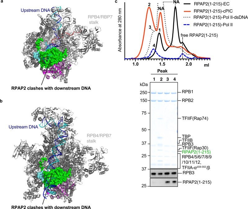
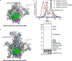
- Fig. 2 RPAP2 binding is incompatible with transcription initiation and elongation. a Superposition of the Pol II-RPAP2 structure onto the Pol II elongation complex (PDB: 5FLM) reveals that RPAP2 would clash with downstream DNA (blue/cyan). b Superposition of the Pol II-RPAP2 structure onto the Pol II pre-initiation complex (PDB: 5IYA) reveals that RPAP2 would clash with downstream DNA (blue/cyan). c Binding competition assays using the preformed Pol II-RPAP2(1-215) complex (peak 4) show that RPAP2 is displaced from Pol II upon formation of an elongation complex (peak 1) or a pre-initiation complex (peak 2) but cannot be displaced by double-stranded promoter DNA (peak 3). Chromatograms show the formation of complexes and the relevant peak fractions used for SDS-PAGE and western blot analysis are indicated. Peaks for free nucleic acids are indicated by NA and vertical dashed lines show the elution peak of free RPAP2(1-215). A representative western blot analysis of the same peak fractions using anti RPAP2 (Thermo Fisher #PA5-61244) and anti RPB3 (BETHYL #A303-771A) antibodies are shown. Please refer to Supplementary Fig. 5 for source data of western blots.