 Explore
 Validate
 Learn
Learn Immunohistochemistry
Antibody data
- Antibody Data
- Antigen structure
- References [1]
- Comments [0]
- Validations
- Immunohistochemistry [5]
- Other assay [4]
Submit
Validation data
Reference
Comment
Report error
- Product number
- PA5-63864 - Provider product page

- Provider
- Invitrogen Antibodies
- Product name
- SLC30A3 Polyclonal Antibody
- Antibody type
- Polyclonal
- Antigen
- Recombinant protein fragment
- Description
- Immunogen sequence: HSHGSRGAEY APLEEGPEEP LPLGNTSVR Highest antigen sequence identity to the following orthologs: Mouse - 79%, Rat - 83%.
- Reactivity
- Human
- Host
- Rabbit
- Isotype
- IgG
- Vial size
- 100 μL
- Concentration
- 0.1 mg/mL
- Storage
- Store at 4°C short term. For long term storage, store at -20°C, avoiding freeze/thaw cycles.
Submitted references Epigenetic targeting of SLC30A3 by HDAC1 is related to the malignant phenotype of glioblastoma.
Zhang L, Liu Z, Dong Y, Kong L
IUBMB life 2021 May;73(5):784-799
IUBMB life 2021 May;73(5):784-799
No comments: Submit comment
Supportive validation
- Submitted by
- Invitrogen Antibodies (provider)
- Main image
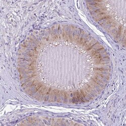
- Experimental details
- Immunohistochemical analysis of SLC30A3 in human epididymis using SLC30A3 Polyclonal Antibody (Product # PA5-63864) shows moderate cytoplasmic positivity in glandular cells.
- Submitted by
- Invitrogen Antibodies (provider)
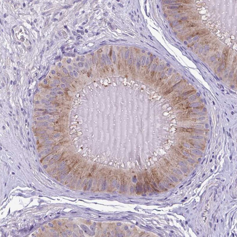
- Main image
- Experimental details
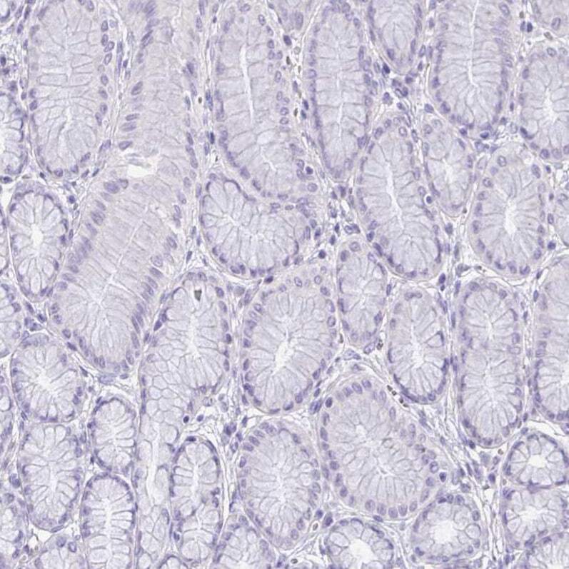
- Immunohistochemical staining of SLC30A3 in human epididymis using SLC30A3 Polyclonal Antibody (Product # PA5-63864) shows high expression.

- Submitted by
- Invitrogen Antibodies (provider)
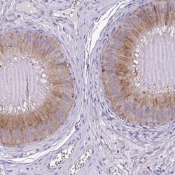
- Main image
- Experimental details
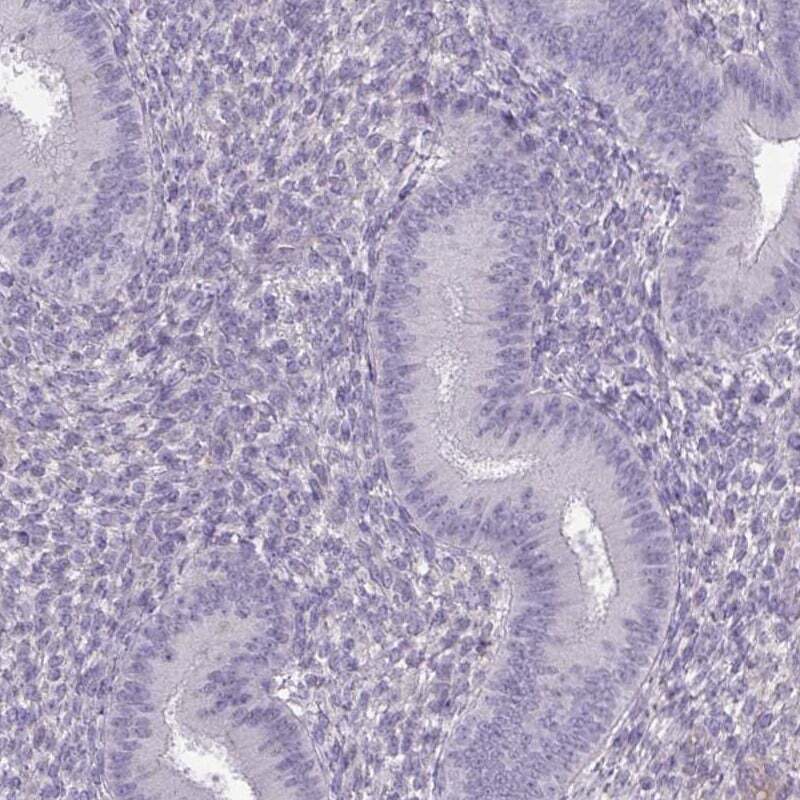
- Immunohistochemical staining of SLC30A3 in human endometrium using SLC30A3 Polyclonal Antibody (Product # PA5-63864) shows low expression as expected.
- Submitted by
- Invitrogen Antibodies (provider)
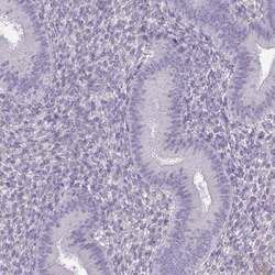
- Main image
- Experimental details
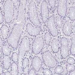
- Immunohistochemical analysis of SLC30A3 in human stomach using SLC30A3 Polyclonal Antibody (Product # PA5-63864) shows no positivity in glandular cells as expected.
- Submitted by
- Invitrogen Antibodies (provider)
- Main image

- Experimental details
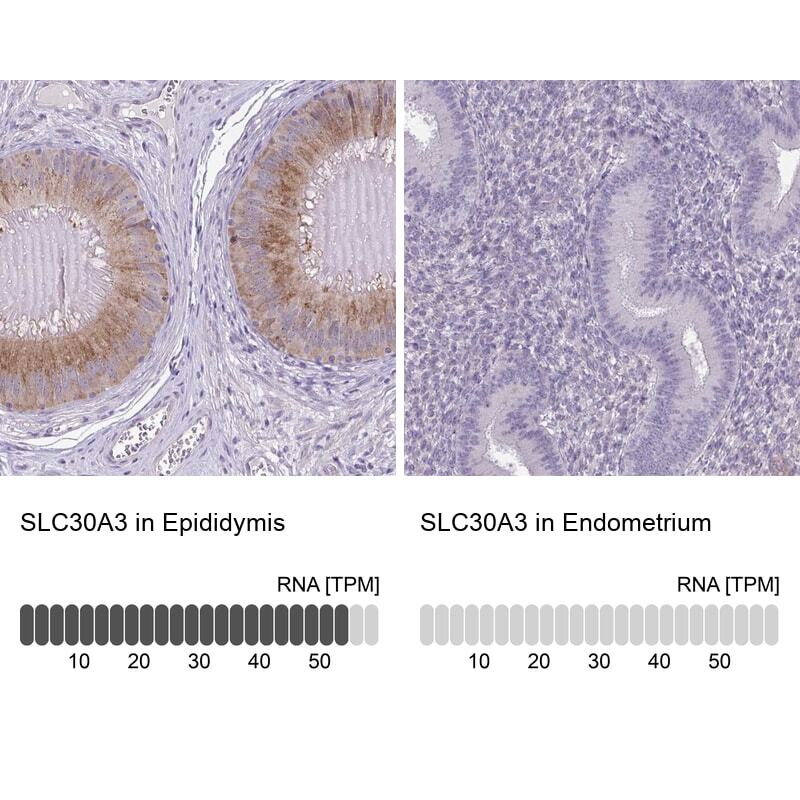
- Immunohistochemical staining of SLC30A3 in human epididymis and endometrium tissues using SLC30A3 Polyclonal Antibody (Product # PA5-63864). Corresponding SLC30A3 RNA-seq data are presented for the same tissues.
Supportive validation
- Submitted by
- Invitrogen Antibodies (provider)
- Main image
- Experimental details
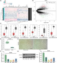
- 1 FIGURE SLC30A3 is diminished in GBM tissues and cell lines. (a) The heatmap of differentially expressed genes in the GSE109857 microarray; (b) the volcanic map of differentially expressed genes in the GSE109857 microarray; (c) expression of SLC30A3, SLC8A2, ITPKA, CRH, CABP1, and KCNJ4 in the TCGA-GBM database; (d) RT-qPCR for SLC30A3 expression in clinically collected cancer tissues and corresponding paracancerous tissues from 47 GBM patients; (e) ISH staining for SLC30A3 distribution in six paired cancerous and paracancerous tissues; (f) RT-qPCR for mRNA expression of SLC30A3 in GBM cells and normal glial cells; (g) western blot for protein expression of SLC30A3 in GBM cells and normal glial cells. Bar indicates means +- SD of three independent experiments. The significance of the differences between two groups was analyzed with paired t test (panel D and E), while the differences among three or more groups was analyzed by using one-way analysis of variance, along with Tukey's test for multiple comparison (panels f and g). * p < .05, *** p < .001
- Submitted by
- Invitrogen Antibodies (provider)
- Main image

- Experimental details
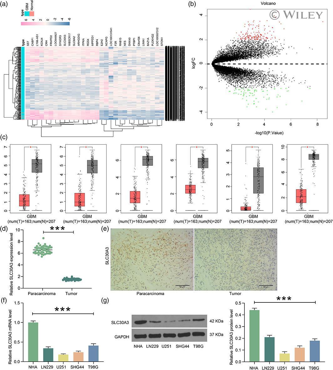
- 2 FIGURE Overexpression of SLC30A3 promotes GBM cell cycle arrest and apoptosis. GBM cells were transfected with SLC30A3-OV or Vector NC. (a) RT-qPCR detection of mRNA expression of SLC30A3 in U251 and SHG44 cells; (b) western blot detection of protein expression of SLC30A3 in U251 and SHG44 cells; (c) CCK-8 detection of U251MG and SHG44 cell activity; (d) flow cytometric analysis of apoptosis levels in U251MG and SHG44 cells; (e) cell cycle distribution in U251 and SHG44 cells detected by flow cytometry; (f) western blot detection of Bcl-2/Bax ratio in U251 and SHG44 cells; (g) western blot detection of Cleaved PARP1 and PUMA expression in U251 and SHG44 cells. Bar indicates means +- SD of three or more independent experiments. The differences among three or more groups were analyzed by using two-way analysis of variance, followed by Tukey's test. ** p < .01
- Submitted by
- Invitrogen Antibodies (provider)
- Main image
- Experimental details
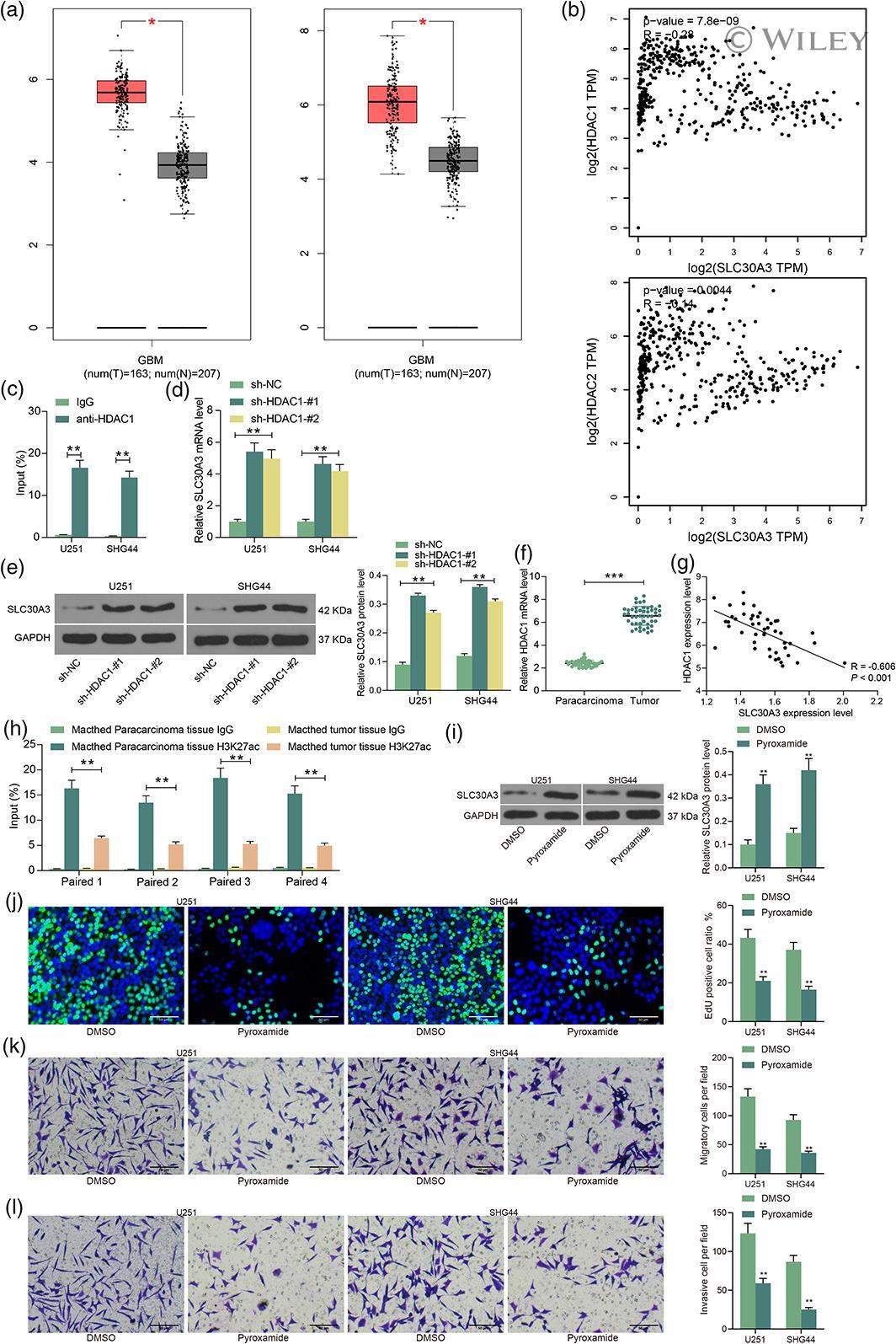
- 6 FIGURE Overexpression of HDAC1 inhibits the expression of SLC30A3 in GBM cells. (a) HDAC1 and HDAC2 expressions were analyzed in the TCGA-GBM database; (b) correlations of HDAC1 and HDAC2 expressions with SLC30A3 expression in the TCGA-GBM database; (c) ChIP-qPCR validation of the binding of HDAC1 to the SE region of SLC30A3; (d) RT-qPCR detection of mRNA expression of SLC30A3 in U251 and SHG44 cells in response to HDAC1 knockdown; (e) western blot detection of protein expression of SLC30A3 in U251 and SHG44 cells in response to HDAC1 knockdown; (f) RT-qPCR for HDAC1 expression in cancerous tissues and corresponding paracancerous tissues of 47 GBM patients collected in clinical trials; (g) Spearman correlation analysis of HDAC1 and SLC30A3 expression in 47 GBM patients; (h) ChIP-qPCR detection of H3K27ac modification of SLC30A3 in four paired cancer tissues and paracancerous tissues. SHG44 and U251 cells were treated with the HDAC1-specific inhibitor Pyroxamide (20.0 muM). (i) Western blot detection of protein expression of SLC30A3 in U251 and SHG44 cells in response to Pyroxamide; (j) EdU detection of U251MG and SHG44 cell activity; (k) Transwell assay detection of migratory capacity of U251 and SHG44 cells; (l) Transwell assay detection of invasive capacity of U251 and SHG44 cells. Bar indicates means +- SD of three independent experiments. The significance of the differences between two groups was analyzed with paired t test (panel f), while the differences among three o
- Submitted by
- Invitrogen Antibodies (provider)
- Main image
- Experimental details
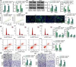
- 7 FIGURE HDAC1 expedites the malignant biological behavior of GBM cells. GBM cells were co-transfected with SLC30A3-OV + HDAC1 or HDAC1 + Vector NC. (a) RT-qPCR detection of mRNA expression of HDAC1 and SLC30A3 in U251 and SHG44 cells; (b) western blot detection of protein expression of HDAC1 and SLC30A3 in U251 and SHG44 cells; (c) CCK-8 detection of U251MG and SHG44 cell proliferation; (d) EdU detection of U251MG and SHG44 cell activity; (e) cell cycle distribution in U251 and SHG44 cells detected by flow cytometry; (f) flow cytometric analysis of apoptosis levels in U251MG and SHG44 cells; (g) Transwell assay detection of migratory capacity of U251 and SHG44 cells; (h) Transwell assay detection of invasive capacity of U251 and SHG44 cells. Bar indicates means +- SD of three independent experiments. The differences among three or more groups were analyzed by using two-way analysis of variance, followed by Tukey's test. ** p < .01
