Explore
 Validate
 Learn
Learn Western blot
 Other assay
Antibody data
- Antibody Data
- Antigen structure
- References [1]
- Comments [0]
- Validations
- Other assay [1]
Submit
Validation data
Reference
Comment
Report error
- Product number
- PA5-71086 - Provider product page

- Provider
- Invitrogen Antibodies
- Product name
- EML5 Polyclonal Antibody
- Antibody type
- Polyclonal
- Antigen
- Synthetic peptide
- Description
- This target displays homology in the following species: Cow: 100%; Dog: 100%; Guinea Pig: 100%; Horse: 100%; Human: 100%; Mouse: 100%; Rabbit: 100%; Rat: 100%; Zebrafish: 100%
- Reactivity
- Human
- Host
- Rabbit
- Isotype
- IgG
- Vial size
- 100 μL
- Concentration
- 0.5 mg/mL
- Storage
- -20°C, Avoid Freeze/Thaw Cycles
Submitted references A Non-Synonymous Point Mutation in a WD-40 Domain Repeat of EML5 Leads to Decreased Bovine Sperm Quality and Fertility.
Nogueira E, Tirpák F, Hamilton LE, Zigo M, Kerns K, Sutovsky M, Kim J, Volkmann D, Jovine L, Taylor JF, Schnabel RD, Sutovsky P
Frontiers in cell and developmental biology 2022;10:872740
Frontiers in cell and developmental biology 2022;10:872740
No comments: Submit comment
Supportive validation
- Submitted by
- Invitrogen Antibodies (provider)
- Main image
- Experimental details
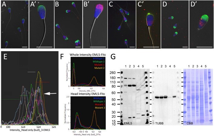
- FIGURE 2 Immunolocalization of EML5 protein (red) in spermatozoa of wild type, heterozygous and homozygous EML5 R1654W bulls. In all immunocytochemistry images acrosomes are stained with PNA (green) and nuclei are stained with DAPI (blue). Panel (A,A') are typical labeling patterns of EML5 in the spermatozoa of wild-type bulls shown at two magnifications. Panel (B) shows EML5 labelling in a heterozygous EML5 R1654W bull (UMC49060) with the increased retention of EML5 in the piriform sperm heads highlighted in (B') . Panel (C) shows the labelling of EML5 in the homozygous EML5 R1654W mutant bull (UMC837), with a single spermatozoon shown at increased magnification in (C') . Images in (D,D') show EML5 labelling in an asthenoteratozoospermic stump tail bull that has a suspected centriolar/microtubular defect but is wildtype for the EML5 mutation. Panel (E) is an image-based flow cytometric histogram overlap of EML5 fluorescence in bulls of varied but acceptable fertility. Each color/curve represents one sire. Intensities of EML5 induced fluorescence are gated from lowest to highest, left to right. The arrow points to the histogram of the heterozygous bull shown in panel (B) , with the highest median value of EML5-induced fluorescence. Panel (F) shows two image-based flow cytometric histograms showing EML5 fluorescence intensity in the whole cell and the isolated head region in two wildtype animals, one heterozygous EML5 R1654W bull (Mutant 1), and one homozygous EML5 R1654W bull