 Explore
 Validate
 Learn
LearnMA5-31801
antibody from Invitrogen Antibodies
Targeting: MOB1A
C2orf6, FLJ10788, FLJ11595, Mats1, MOB1, Mob4B, MOBK1B, MOBKL1B
 Western blot
 ELISA
ELISAAntibody data
- Antibody Data
- Antigen structure
- References [1]
- Comments [0]
- Validations
- ELISA [2]
- Flow cytometry [2]
- Other assay [1]
Submit
Validation data
Reference
Comment
Report error
- Product number
- MA5-31801 - Provider product page

- Provider
- Invitrogen Antibodies
- Product name
- MOB1A Monoclonal Antibody (3E7B2)
- Antibody type
- Monoclonal
- Antigen
- Purifed from natural sources
- Description
- MA5-31801 has been tested in indirect ELISA.
- Reactivity
- Human
- Host
- Mouse
- Isotype
- IgG
- Antibody clone number
- 3E7B2
- Vial size
- 100 μL
- Concentration
- 1 mg/mL
- Storage
- Store at 4°C short term. For long term storage, store at -20°C, avoiding freeze/thaw cycles.
Submitted references Thrombospondin-1 Plays an Essential Role in Yes-Associated Protein Nuclear Translocation during the Early Phase of Trypanosoma cruzi Infection in Heart Endothelial Cells.
Arun A, Rayford KJ, Cooley A, Rachakonda G, Villalta F, Pratap S, Lima MF, Sheibani N, Nde PN
International journal of molecular sciences 2020 Jul 12;21(14)
International journal of molecular sciences 2020 Jul 12;21(14)
No comments: Submit comment
Supportive validation
- Submitted by
- Invitrogen Antibodies (provider)
- Main image

- Experimental details
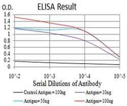
- ELISA analysis of MOB1 in Control Antigen (black line, 100 ng); Antigen (purple line, 10 ng); Antigen (blue line, 50 ng); Antigen (red line, 100 ng). Samples were incubated with MOB1 monoclonal antibody (Product # MA5-31801) using a dilution of 1:10,000.

- Submitted by
- Invitrogen Antibodies (provider)
- Main image
- Experimental details
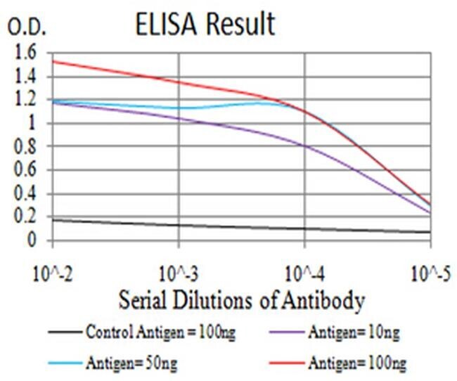
- ELISA analysis of MOB1 in Control Antigen (black line, 100 ng); Antigen (purple line, 10 ng); Antigen (blue line, 50 ng); Antigen (red line, 100 ng). Samples were incubated with MOB1 monoclonal antibody (Product # MA5-31801) using a dilution of 1:10,000.
Supportive validation
- Submitted by
- Invitrogen Antibodies (provider)
- Main image

- Experimental details
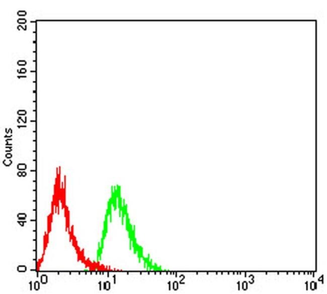
- Flow cytometry of MOB1 in HeLa cells (green). Sample was incubated with MOB1 monoclonal antibody (Product # MA5-31801) using a dilution of 1:200-1:400 followed by negative control (red).

- Submitted by
- Invitrogen Antibodies (provider)
- Main image

- Experimental details
- Flow cytometry of MOB1 in HeLa cells (green). Sample was incubated with MOB1 monoclonal antibody (Product # MA5-31801) using a dilution of 1:200-1:400 followed by negative control (red).
Supportive validation
- Submitted by
- Invitrogen Antibodies (provider)
- Main image

- Experimental details
- Figure 1 TSP-1 is essential for activation of hippo signaling cascade during the early phase of T. cruzi infection. Lysates (20 µg) from WT or TSP-1KO MHEC challenged with T . cruzi at different time points were resolved by SDS-PAGE, blotted, and probed with antibodies against ( A ) SAV1, ( B ) MOB1A in WT MHEC and ( C ) SAV1, ( D ) MOB1A in TSP-1KO MHEC, and developed as described. The blots were stripped, reprobed with antibodies against housekeeping GAPDH and developed with the corresponding IRDye conjugated secondary antibody. The developed blots were scanned using the infrared fluorescence detection Odyssey Imaging Systems. The normalized fold change in the level of each unphosphorylated protein was determined and plotted in the bar graph for WT MHEC ( A , lower panel) SAV1, ( B , lower panel) MOB1A, respectively and for TSP-1KO MHEC ( C , lower panel) SAV1 and ( D , lower panel) MOB1A, respectively. The bar graphs represent mean values +- SE from three independent biological replicates. The value of p < 0.05 was considered significant. * p < 0.05; ** p < 0.01; *** p < 0.001.
