Explore
 Validate
 Learn
Learn Western blot
 Immunocytochemistry
Antibody data
- Antibody Data
- Antigen structure
- References [2]
- Comments [0]
- Validations
- Immunocytochemistry [1]
- Immunohistochemistry [2]
Submit
Validation data
Reference
Comment
Report error
- Product number
- HPA019687 - Provider product page

- Provider
- Atlas Antibodies
- Proper citation
- Atlas Antibodies Cat#HPA019687, RRID:AB_1857980
- Product name
- Anti-THOC1
- Antibody type
- Polyclonal
- Description
- Polyclonal Antibody against Human THOC1, Gene description: THO complex 1, Alternative Gene Names: HPR1, P84, Validated applications: IHC, ICC, WB, Uniprot ID: Q96FV9, Storage: Store at +4°C for short term storage. Long time storage is recommended at -20°C.
- Reactivity
- Human, Mouse, Rat
- Host
- Rabbit
- Conjugate
- Unconjugated
- Isotype
- IgG
- Vial size
- 100 µl
- Concentration
- 0.4 mg/ml
- Storage
- Store at +4°C for short term storage. Long time storage is recommended at -20°C.
- Handling
- The antibody solution should be gently mixed before use.
Submitted references The HIF transcription network exerts innate antiviral activity in neurons and limits brain inflammation
Differential expression of THOC1 and ALY mRNP biogenesis/export factors in human cancers
Farahani E, Reinert L, Narita R, Serrero M, Skouboe M, van der Horst D, Assil S, Zhang B, Iversen M, Gutierrez E, Hazrati H, Johannsen M, Olagnier D, Kunze R, Denham M, Mogensen T, Lappe M, Paludan S
Cell Reports 2024;43(2):113792
Cell Reports 2024;43(2):113792
Differential expression of THOC1 and ALY mRNP biogenesis/export factors in human cancers
Domínguez-Sánchez M, Sáez C, Japón M, Aguilera A, Luna R
BMC Cancer 2011;11(1)
BMC Cancer 2011;11(1)
No comments: Submit comment
Supportive validation
- Submitted by
- Atlas Antibodies (provider)
- Main image
- Experimental details
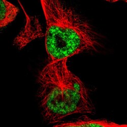
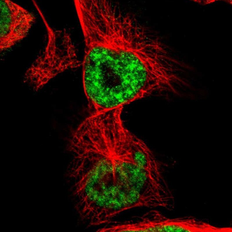
- Immunofluorescent staining of human cell line U-251 MG shows localization to nuclear speckles.
- Sample type
- Human
Supportive validation
Supportive validation
- Submitted by
- Atlas Antibodies (provider)
- Enhanced method
- Orthogonal validation
- Main image
- Experimental details
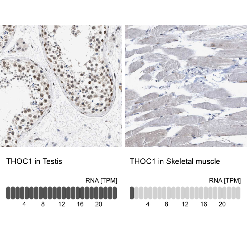
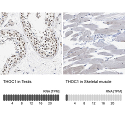
- Immunohistochemistry analysis in human testis and skeletal muscle tissues using Anti-THOC1 antibody. Corresponding THOC1 RNA-seq data are presented for the same tissues.
- Sample type
- Human
- Protocol
- Protocol

Supportive validation
- Submitted by
- Atlas Antibodies (provider)
- Enhanced method
- Orthogonal validation
- Main image
- Experimental details
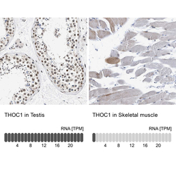
- Immunohistochemistry analysis in human testis and skeletal muscle tissues using Anti-THOC1 antibody. Corresponding THOC1 RNA-seq data are presented for the same tissues.
- Sample type
- Human
- Protocol
- Protocol