 Explore
 Validate
 Learn
LearnMA1-23261
antibody from Invitrogen Antibodies
Targeting: THOC1
HPR1, P84
 Western blot
 Immunocytochemistry Immunoprecipitation Immunohistochemistry
 Chromatin Immunoprecipitation Other assay
Antibody data
- Antibody Data
- Antigen structure
- References [1]
- Comments [0]
- Validations
- Immunocytochemistry [4]
- Immunoprecipitation [1]
- Immunohistochemistry [2]
- Other assay [1]
Submit
Validation data
Reference
Comment
Report error
- Product number
- MA1-23261 - Provider product page

- Provider
- Invitrogen Antibodies
- Product name
- Nuclear Matrix Protein p84 Monoclonal Antibody (5E10)
- Antibody type
- Monoclonal
- Antigen
- Other
- Description
- Suggested positive controls are HeLa, Raji, and MOLT4.
- Reactivity
- Human, Mouse, Rat, Hamster
- Host
- Mouse
- Isotype
- IgG
- Antibody clone number
- 5E10
- Vial size
- 100 μL
- Concentration
- 1 mg/mL
- Storage
- Store at 4°C short term. For long term storage, store at -20°C, avoiding freeze/thaw cycles.
Submitted references Dihydroartemisinin inhibits the viability of cervical cancer cells by upregulating caveolin 1 and mitochondrial carrier homolog 2: Involvement of p53 activation and NAD(P)H:quinone oxidoreductase 1 downregulation.
Zhang T, Hu Y, Wang T, Cai P
International journal of molecular medicine 2017 Jul;40(1):21-30
International journal of molecular medicine 2017 Jul;40(1):21-30
No comments: Submit comment
Supportive validation
- Submitted by
- Invitrogen Antibodies (provider)
- Main image
- Experimental details
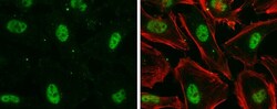
- Immunocytochemistry-Immunofluorescence analysis of Nuclear Matrix Protein p84 was performed in HeLa cells fixed in 4% paraformaldehyde at RT for 15 min. Green: Nuclear Matrix Protein p84 Monoclonal Antibody (5E10) (Product # MA1 23261) diluted at 1:500. Red: phalloidin, a cytoskeleton marker.

- Submitted by
- Invitrogen Antibodies (provider)
- Main image

- Experimental details
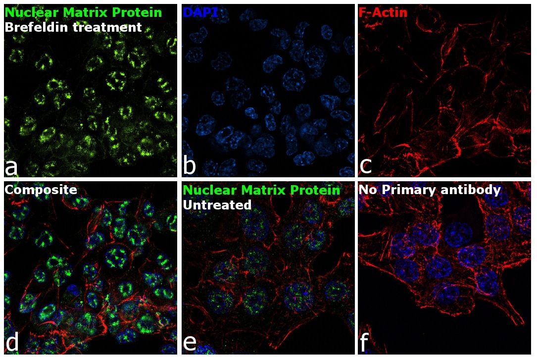
- Immunofluorescence analysis of Nuclear Matrix Protein p84 was performed using 70% confluent log phase HCT 116 cells treated with 0.5 µg of Brefeldin for 24 hours. The cells were fixed with 4% paraformaldehyde for 10 minutes, permeabilized with 0.1% Triton™ X-100 for 15 minutes, and blocked with 1% BSA for 1 hour at room temperature. The cells were labeled with Nuclear Matrix Protein p84 Mouse Monoclonal Antibody (Product # MA1-23261) at 5 µg/mL in 0.1% BSA, incubated at 4 degree Celsius overnight and then labeled with Goat anti-Mouse IgG (H+L) Superclonal™ Secondary Antibody, Alexa Fluor® 488 conjugate (Product # A28175) at a dilution of 1:2000 for 45 minutes at room temperature (Panel a: green). Nuclei (Panel b: blue) were stained with ProLong™ Diamond Antifade Mountant with DAPI (Product # P36962). F-actin (Panel c: red) was stained with Rhodamine Phalloidin (Product # R415, 1:300). Panel d represents the merged image showing Nuclear localization. Panel e shows untreated cells with less Nuclear signal. Panel f represents control cells with no primary antibody to assess background. The images were captured at 60X magnification.
- Submitted by
- Invitrogen Antibodies (provider)
- Main image

- Experimental details
- Immunocytochemistry-Immunofluorescence analysis of Nuclear Matrix Protein p84 was performed in HeLa cells fixed in 4% paraformaldehyde at RT for 15 min. Green: Nuclear Matrix Protein p84 Monoclonal Antibody (5E10) (Product # MA1 23261) diluted at 1:500. Red: phalloidin, a cytoskeleton marker.

- Submitted by
- Invitrogen Antibodies (provider)
- Main image

- Experimental details
- Immunofluorescence analysis of Nuclear Matrix Protein p84 was performed using 70% confluent log phase HCT 116 cells treated with 0.5 µg of Brefeldin for 24 hours. The cells were fixed with 4% paraformaldehyde for 10 minutes, permeabilized with 0.1% Triton™ X-100 for 15 minutes, and blocked with 1% BSA for 1 hour at room temperature. The cells were labeled with Nuclear Matrix Protein p84 Mouse Monoclonal Antibody (Product # MA1-23261) at 5 µg/mL in 0.1% BSA, incubated at 4 degree Celsius overnight and then labeled with Goat anti-Mouse IgG (H+L) Superclonal™ Secondary Antibody, Alexa Fluor® 488 conjugate (Product # A28175) at a dilution of 1:2000 for 45 minutes at room temperature (Panel a: green). Nuclei (Panel b: blue) were stained with ProLong™ Diamond Antifade Mountant with DAPI (Product # P36962). F-actin (Panel c: red) was stained with Rhodamine Phalloidin (Product # R415, 1:300). Panel d represents the merged image showing Nuclear localization. Panel e shows untreated cells with less Nuclear signal. Panel f represents control cells with no primary antibody to assess background. The images were captured at 60X magnification.

Supportive validation
- Submitted by
- Invitrogen Antibodies (provider)
- Main image

- Experimental details
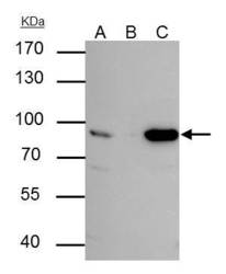
- p84 antibody [5E10] immunoprecipitates p84 protein in IP experiments. IP Sample: HepG2 whole cell lysate/extract A : 30 µg whole cell lysate/extract of p84 protein expressing HepG2 cells B : Control with 3 µg of pre-immune mouse IgG C : Immunoprecipitation of p84 by 3 µg of p84 antibody [5E10] (Product # MA1-23261) 7.5% SDS-PAGE The immunoprecipitated p84 protein was detected by p84 antibody [5E10] (Product # MA1-23261) diluted at 1 : 1,000.

Supportive validation
- Submitted by
- Invitrogen Antibodies (provider)
- Main image
- Experimental details
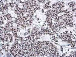
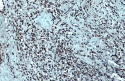
- Immunohistochemistry (Paraffin) analysis of Nuclear Matrix Protein p84 was performed in paraffin-embedded human breast carcinoma tissue using Nuclear Matrix Protein p84 Monoclonal Antibody (5E10) (Product # MA1-23261) at a dilution of 1:200. Antigen Retrieval: Citrate buffer, pH 6.0, 15 min.
- Submitted by
- Invitrogen Antibodies (provider)
- Main image
- Experimental details
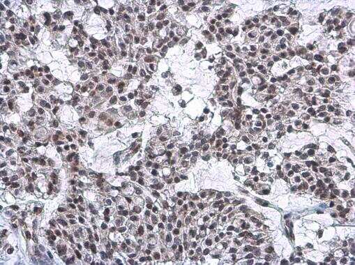
- Immunohistochemistry (Paraffin) analysis of Nuclear Matrix Protein p84 was performed in paraffin-embedded human lung cancer tissue using Nuclear Matrix Protein p84 Monoclonal Antibody (5E10) (Product # MA1-23261) at a dilution of 1:100. Antigen Retrieval: Citrate buffer, pH 6.0, 15 min.

Supportive validation
- Submitted by
- Invitrogen Antibodies (provider)
- Main image
- Experimental details
- p84 antibody [5E10] immunoprecipitates p84 protein in IP experiments. IP Sample: HepG2 whole cell lysate/extract A : 30 µg whole cell lysate/extract of p84 protein expressing HepG2 cells B : Control with 3 µg of pre-immune mouse IgG C : Immunoprecipitation of p84 by 3 µg of p84 antibody [5E10] (Product # MA1-23261) 7.5% SDS-PAGE The immunoprecipitated p84 protein was detected by p84 antibody [5E10] (Product # MA1-23261) diluted at 1 : 1,000.
