Explore
 Validate
 Learn
LearnPA5-29316
antibody from Invitrogen Antibodies
Targeting: DDR1
CAK, CD167, EDDR1, NEP, NTRK4, PTK3A, RTK6
 Western blot
 Immunohistochemistry
Antibody data
- Antibody Data
- Antigen structure
- References [1]
- Comments [0]
- Validations
- Immunohistochemistry [1]
- Other assay [1]
Submit
Validation data
Reference
Comment
Report error
- Product number
- PA5-29316 - Provider product page

- Provider
- Invitrogen Antibodies
- Product name
- MCK10 Polyclonal Antibody
- Antibody type
- Polyclonal
- Antigen
- Recombinant full-length protein
- Description
- Recommended positive controls: A431, H1299, HeLa, HepG2, Raji, mouse brain. Predicted reactivity: Mouse (89%), Rat (91%), Pig (93%), Rhesus Monkey (99%), Chimpanzee (100%), Bovine (91%). Store product as a concentrated solution. Centrifuge briefly prior to opening the vial.
- Reactivity
- Human, Mouse
- Host
- Rabbit
- Isotype
- IgG
- Vial size
- 100 μL
- Concentration
- 1 mg/mL
- Storage
- Store at 4°C short term. For long term storage, store at -20°C, avoiding freeze/thaw cycles.
Submitted references Discoidin Domain Receptor 1 Regulates Runx2 during Osteogenesis of Osteoblasts and Promotes Bone Ossification via Phosphorylation of p38.
Chou LY, Chen CH, Chuang SC, Cheng TL, Lin YH, Chou HC, Fu YC, Wang YH, Wang CZ
International journal of molecular sciences 2020 Sep 29;21(19)
International journal of molecular sciences 2020 Sep 29;21(19)
No comments: Submit comment
Supportive validation
- Submitted by
- Invitrogen Antibodies (provider)
- Main image

- Experimental details
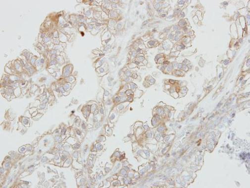
- Immunohistochemical analysis of paraffin-embedded human ovarian cancer, using DDR1 (Product # PA5-29316) antibody at 1:500 dilution. Antigen Retrieval: EDTA based buffer, pH 8.0, 15 min.
Supportive validation
- Submitted by
- Invitrogen Antibodies (provider)
- Main image
- Experimental details
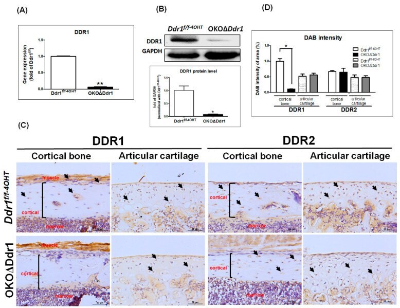
- Figure 1 Generation of osteoblast-specific (a1(I) collagen-CreERT; Ddr1 f/f ) OKODelta Ddr1 mice. The calvarial bone was extracted from Ddr1 f/f-4OHT and OKODelta Ddr1 mice on postnatal days 4~5, and ( A ) gene expression of Ddr1 and ( B ) protein level of DDR1 were detected. The ratio of DDR1/GPADH is expressed relative to the Ddr1 f/f-4OHT mice was quantified. Each group n >= 6. ( C ) IHC staining of DDR1 and DDR2 in cortical bone and articular cartilage of femurs from 4-week-old mice; black arrow indicates DDR1-positive cells; black frame indicates the cortical region. ( D ) Quantitative results by tissue faxes normalized with Ddr1 f/f-4OHT . Magnifications of 400X are shown, with scale bars of 50 mum. Each group, n >= 6; * p