Explore
 Validate
 Learn
Learn Western blot
Antibody data
- Antibody Data
- Antigen structure
- References [3]
- Comments [0]
- Validations
- Western blot [2]
- Immunohistochemistry [1]
Submit
Validation data
Reference
Comment
Report error
- Product number
- AF2396 - Provider product page

- Provider
- R&D Systems
- Product name
- Human DDR1 Antibody
- Antibody type
- Polyclonal
- Description
- Immunogen affinity purified. Detects human DDR1 in direct ELISAs and Western blots. In sandwich immunoassays, approximately 5% cross-reactivity with recombinant human DDR2 is observed.
- Reactivity
- Human
- Host
- Goat
- Conjugate
- Unconjugated
- Antigen sequence
Q08345- Isotype
- IgG
- Vial size
- 100 ug
- Concentration
- LYOPH
- Storage
- Use a manual defrost freezer and avoid repeated freeze-thaw cycles. 12 months from date of receipt, -20 to -70 °C as supplied. 1 month, 2 to 8 °C under sterile conditions after reconstitution. 6 months, -20 to -70 °C under sterile conditions after reconstitution.
Submitted references Cancer Cell Invasion in Three-dimensional Collagen Is Regulated Differentially by Gα13 Protein and Discoidin Domain Receptor 1-Par3 Protein Signaling.
Shedding of discoidin domain receptor 1 by membrane-type matrix metalloproteinases.
Type II collagen levels correlate with mineralization by articular cartilage vesicles.
Chow CR, Ebine K, Knab LM, Bentrem DJ, Kumar K, Munshi HG
The Journal of biological chemistry 2016 Jan 22;291(4):1605-1618
The Journal of biological chemistry 2016 Jan 22;291(4):1605-1618
Shedding of discoidin domain receptor 1 by membrane-type matrix metalloproteinases.
Fu HL, Sohail A, Valiathan RR, Wasinski BD, Kumarasiri M, Mahasenan KV, Bernardo MM, Tokmina-Roszyk D, Fields GB, Mobashery S, Fridman R
The Journal of biological chemistry 2013 Apr 26;288(17):12114-29
The Journal of biological chemistry 2013 Apr 26;288(17):12114-29
Type II collagen levels correlate with mineralization by articular cartilage vesicles.
Jubeck B, Muth E, Gohr CM, Rosenthal AK
Arthritis and rheumatism 2009 Sep;60(9):2741-6
Arthritis and rheumatism 2009 Sep;60(9):2741-6
No comments: Submit comment
Supportive validation
- Submitted by
- R&D Systems (provider)
- Main image
- Experimental details
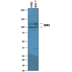
- Detection of Human DDR1 by Western Blot. Western blot shows lysates of K562 human chronic myelogenous leukemia cell line and MCF-7 human breast cancer cell line. PVDF membrane was probed with 1 µg/mL of Goat Anti-Human DDR1 Antigen Affinity-purified Polyclonal Antibody (Catalog # AF2396) followed by HRP-conjugated Anti-Goat IgG Secondary Antibody (Catalog # HAF019). Specific bands were detected for DDR1 at approximately 100 and 120 kDa (as indicated). This experiment was conducted under reducing conditions and using Immunoblot Buffer Group 1.

- Submitted by
- R&D Systems (provider)
- Main image
- Experimental details
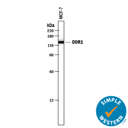
- Detection of Human DDR1 by Simple WesternTM. Simple Western lane view shows lysates of MCF-7 human breast cancer cell line, loaded at 0.2 mg/mL. A specific band was detected for DDR1 at approximately 137 kDa (as indicated) using 10 µg/mL of Goat Anti-Human DDR1 Antigen Affinity-purified Polyclonal Antibody (Catalog # AF2396) followed by 1:50 dilution of HRP-conjugated Anti-Goat IgG Secondary Antibody (Catalog # HAF109). This experiment was conducted under reducing conditions and using the 12-230 kDa separation system.

Supportive validation
- Submitted by
- R&D Systems (provider)
- Main image
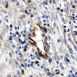
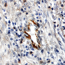
- Experimental details
- DDR1 in Human Breast Cancer Tissue. DDR1 was detected in immersion fixed paraffin-embedded sections of human breast cancer tissue using Human DDR1 Antigen Affinity-purified Polyclonal Antibody (Catalog # AF2396) at 15 µg/mL overnight at 4 °C. Before incubation with the primary antibody tissue was subjected to heat-induced epitope retrieval using Antigen Retrieval Reagent-Basic (Catalog # CTS013). Tissue was stained using the Anti-Goat HRP-DAB Cell & Tissue Staining Kit (brown; Catalog # CTS008) and counterstained with hematoxylin (blue). View our protocol for Chromogenic IHC Staining of Paraffin-embedded Tissue Sections.