Explore
 Validate
 Learn
Learn Western blot
Antibody data
- Antibody Data
- Antigen structure
- References [0]
- Comments [0]
- Validations
- Western blot [2]
- Immunocytochemistry [1]
- Immunohistochemistry [1]
- Flow cytometry [1]
Submit
Validation data
Reference
Comment
Report error
- Product number
- AP52089PU-N - Provider product page

- Provider
- OriGene
- Product name
- Haptoglobin (HP) (Center) rabbit polyclonal antibody, Aff - Purified
- Antibody type
- Polyclonal
- Description
- Haptoglobin (HP) (Center) rabbit polyclonal antibody, Aff - Purified
- Host
- Rabbit
- Conjugate
- Unconjugated
- Epitope
- HP
- Antibody clone number
- NULL
- Vial size
- 400 µl
- Concentration
- lot specific
No comments: Submit comment
Supportive validation
- Submitted by
- OriGene (provider)
- Main image

- Experimental details
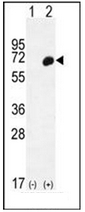
- Western blot analysis of HP (arrow) using Haptoglobin?Antibody (Center) . 293 cell lysates (2 ug/lane) either nontransfected (Lane 1) or transiently transfected (Lane 2) with the HP gene.
- Validation comment
- WB
- Submitted by
- OriGene (provider)
- Main image

- Experimental details
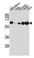
- Western blot analysis of Haptoglobin?Antibody (Center) ??in MDA-MB453,CEM,NCI-H460,HepG2,K562 cell line lysates (35ug/lane). This demonstrates the HP antibody detected the HP protein (arrow).
- Validation comment
- WB
Supportive validation
- Submitted by
- OriGene (provider)
- Main image

- Experimental details
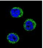
- Confocal immunofluorescent analysis of Haptoglobin?Antibody (Center) ??with MDA-MB435 cell followed by Alexa Fluor 488-conjugated goat anti-rabbit lgG (green).DAPI was used to stain the cell nuclear (blue).
- Validation comment
- IF
Supportive validation
- Submitted by
- OriGene (provider)
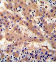
- Main image

- Experimental details
- Immunohistochemistry analysis in formalin fixed and paraffin embedded human hepatocarcinoma reacted with Haptoglobin?Antibody (Center) ??followed by peroxidase conjugation of the secondary antibody and DAB staining.
- Validation comment
- IHC
Supportive validation
- Submitted by
- OriGene (provider)
- Main image

- Experimental details
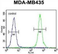
- Flow cytometric analysis of MDA-MB435 cells using Haptoglobin?Antibody (Center) ??(right histogram) compared to a negative control cell (left histogram). FITC-conjugated goat-anti-rabbit secondary antibodies were used for the analysis.
- Validation comment
- FC