Explore
 Validate
 Learn
Learn Western blot
 Immunocytochemistry
Antibody data
- Antibody Data
- Antigen structure
- References [1]
- Comments [0]
- Validations
- Immunocytochemistry [2]
- Immunohistochemistry [2]
- Flow cytometry [2]
- Other assay [1]
Submit
Validation data
Reference
Comment
Report error
- Product number
- PA5-24174 - Provider product page

- Provider
- Invitrogen Antibodies
- Product name
- Haptoglobin Polyclonal Antibody
- Antibody type
- Polyclonal
- Antigen
- Synthetic peptide
- Reactivity
- Human, Mouse
- Host
- Rabbit
- Isotype
- IgG
- Vial size
- 400 μL
- Concentration
- 0.5 mg/mL
- Storage
- Store at 4°C short term. For long term storage, store at -20°C, avoiding freeze/thaw cycles.
Submitted references Hypertransaminasemia and liver fibrosis associated with haptoglobin retention and anhaptoglobinemia in a paediatric patient.
Gunzer S, Kraus A, Buchroth I, Grüneberg M, Westermann C, Biskup S, Reunert J, Grünewald I, Marquardt T
Liver international : official journal of the International Association for the Study of the Liver 2021 Oct;41(10):2427-2432
Liver international : official journal of the International Association for the Study of the Liver 2021 Oct;41(10):2427-2432
No comments: Submit comment
Supportive validation
- Submitted by
- Invitrogen Antibodies (provider)
- Main image
- Experimental details
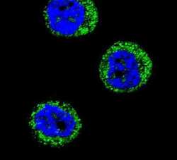
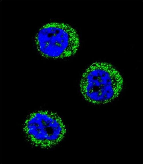
- Immunofluorescent analysis of MDA-MB435 cells using a HP polyclonal antibody (Product # PA5-24174) at a dilution of 1:10-50, followed by a fluor-conjugated goat anti-rabbit secondary antibody (green). Nuclei were stained with DAPI (blue).

- Submitted by
- Invitrogen Antibodies (provider)
- Main image

- Experimental details
- Immunocytochemistry analysis of Haptoglobin in MDA-MB435 cells. Samples were incubated in Haptoglobin polyclonal antibody (Product # PA5-24174) followed by Alexa Fluor 488-conjugated goat anti-rabbit lgG (green). DAPI was used to stain the cell nuclear (blue).
Supportive validation
- Submitted by
- Invitrogen Antibodies (provider)
- Main image
- Experimental details
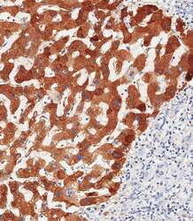
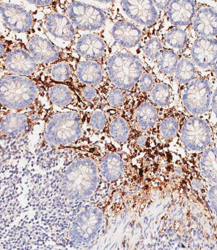
- Immunohistochemistry analysis of Haptoglobin in paraffin-embedded human hepatocarcinoma tissue. Samples were incubated with Haptoglobin polyclonal antibody (Product # PA5-24174) using a dilution of 1:500 for 1 hour at room temperature followed by an undiluted biotinylated CRF Anti-Polyvalent HRP Polymer antibody. Tissue was fixed with formaldehyde at room temperature, antigen retrieval was by heat mediation with a EDTA buffer (pH 9.0).

- Submitted by
- Invitrogen Antibodies (provider)
- Main image

- Experimental details
- Immunohistochemistry analysis of Haptoglobin in paraffin-embedded human colon tissue. Samples were incubated with Haptoglobin polyclonal antibody (Product # PA5-24174) using a dilution of 1:500 for 1 hour at room temperature followed by an undiluted biotinylated CRF Anti-Polyvalent HRP Polymer antibody. Tissue was fixed with formaldehyde at room temperature, antigen retrieval was by heat mediation with a EDTA buffer (pH 9.0).
Supportive validation
- Submitted by
- Invitrogen Antibodies (provider)
- Main image
- Experimental details
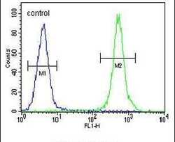
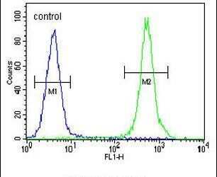
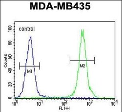
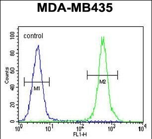
- Flow cytometry analysis of MDA-MB435 cells using a HP polyclonal antibody (Product # PA5-24174) (right) compared to a negative control cell (left) at a dilution of 1:10-50, followed by a FITC-conjugated goat anti-rabbit antibody
- Submitted by
- Invitrogen Antibodies (provider)
- Main image
- Experimental details
- Flow cytometry of Haptoglobin in MDA-MB435 cells (right histogram). Samples were incubated with Haptoglobin polyclonal antibody (Product # PA5-24174) followed by FITC-conjugated goat-anti-rabbit secondary antibody. Negative control cell (left histogram).
Supportive validation
- Submitted by
- Invitrogen Antibodies (provider)
- Main image

- Experimental details
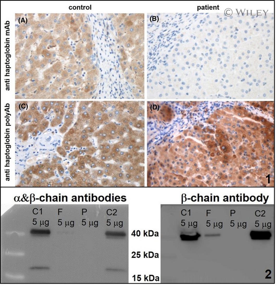
- 2 FIGURE Immunohistochemistry for immunodetection of Hp in liver biopsy tissue and Western Blot. 2.1 Immunohistochemistry for immunodetection of Hp in liver biopsy tissue. A, Control tissue stained with the monoclonal anti-haptoglobin antibody. Positive staining reaction shows (brown) presence of haptoglobin in the cytoplasm of hepatocytes. Original magnification 400x. B, The monoclonal antibody is not able to detect haptoglobin in the patient-derived tissue. Original magnification 400x. C, Control tissue stained with the polyclonal anti-haptoglobin antibody. Positive staining reaction shows presence of haptoglobin in the hepatocytes. Original magnification 400x. D, Patient`s tissue stained with the polyclonal anti-haptoglobin antibody. Positive staining reaction shows presence of haptoglobin in the hepatocytes. Original magnification 400x. 2.2 Western Blot detection of Haptoglobin in blood serum. C1, C2 = control serum of healthy individuals (m/f) show normal level of haptoglobin. F = patient`s father, shows low level of haptoglobin. P = patient shows no serum haptoglobin. alpha-chain (alpha-2) = 18 kDa. beta-chain = 40 kDa