 Explore
 Validate
 Learn
LearnH00001523-M01
antibody from Novus Biologicals
Targeting: CUX1
CASP, CDP, CDP/Cut, CDP/Cux, CDP1, Clox, CUT, CUTL1, CUX, Cux/CDP, GOLIM6
 Western blot
Antibody data
- Antibody Data
- Antigen structure
- References [3]
- Comments [0]
- Validations
- Western blot [1]
- ELISA [1]
- Immunocytochemistry [1]
- Immunoprecipitation [1]
- Immunohistochemistry [2]
Submit
Validation data
Reference
Comment
Report error
- Product number
- H00001523-M01 - Provider product page

- Provider
- Novus Biologicals
- Proper citation
- Novus Cat#H00001523-M01, RRID:AB_538074
- Product name
- Mouse Monoclonal CDP/CUTL1 Antibody
- Antibody type
- Monoclonal
- Description
- IgG purified. CUTL1 - cut-like 1, CCAAT displacement protein (Drosophila)
- Reactivity
- Human, Mouse, Rat
- Host
- Mouse
- Isotype
- IgG
- Vial size
- 0.1 mg
- Storage
- Aliquot and store at -20C or -80C. Avoid freeze-thaw cycles.
Submitted references Developmental and adult expression patterns of the G-protein-coupled receptor GPR88 in the rat: Establishment of a dual nuclear-cytoplasmic localization.
Functional cortical neurons and astrocytes from human pluripotent stem cells in 3D culture.
Neocortical layer formation of human developing brains and lissencephalies: consideration of layer-specific marker expression.
Massart R, Mignon V, Stanic J, Munoz-Tello P, Becker JA, Kieffer BL, Darmon M, Sokoloff P, Diaz J
The Journal of comparative neurology 2016 Oct 1;524(14):2776-802
The Journal of comparative neurology 2016 Oct 1;524(14):2776-802
Functional cortical neurons and astrocytes from human pluripotent stem cells in 3D culture.
Paşca AM, Sloan SA, Clarke LE, Tian Y, Makinson CD, Huber N, Kim CH, Park JY, O'Rourke NA, Nguyen KD, Smith SJ, Huguenard JR, Geschwind DH, Barres BA, Paşca SP
Nature methods 2015 Jul;12(7):671-8
Nature methods 2015 Jul;12(7):671-8
Neocortical layer formation of human developing brains and lissencephalies: consideration of layer-specific marker expression.
Saito T, Hanai S, Takashima S, Nakagawa E, Okazaki S, Inoue T, Miyata R, Hoshino K, Akashi T, Sasaki M, Goto Y, Hayashi M, Itoh M
Cerebral cortex (New York, N.Y. : 1991) 2011 Mar;21(3):588-96
Cerebral cortex (New York, N.Y. : 1991) 2011 Mar;21(3):588-96
No comments: Submit comment
Supportive validation
- Submitted by
- Novus Biologicals (provider)
- Main image
- Experimental details
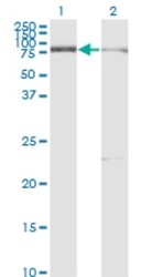
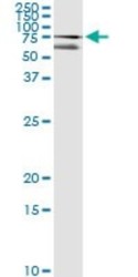
- Western Blot: CDP/CUTL1 Antibody (2A10) [H00001523-M01] - Analysis of CUX1 expression in transfected 293T cell line by CUTL1 monoclonal antibody (M01), clone 2A10.Lane 1: CUX1 transfected lysate(77.2 KDa).Lane 2: Non-transfected lysate.
Supportive validation
- Submitted by
- Novus Biologicals (provider)
- Main image
- Experimental details
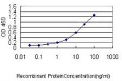
- ELISA: CDP/CUTL1 Antibody (2A10) [H00001523-M01] - Detection limit for recombinant GST tagged CUTL1 is approximately 0.03ng/ml as a capture antibody.
Supportive validation
- Submitted by
- Novus Biologicals (provider)
- Main image

- Experimental details
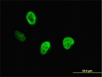
- Immunocytochemistry/Immunofluorescence: CDP/CUTL1 Antibody (2A10) [H00001523-M01] - Analysis of monoclonal antibody to CUTL1 on HeLa cell. Antibody concentration 10 ug/ml.
Supportive validation
- Submitted by
- Novus Biologicals (provider)
- Main image
- Experimental details
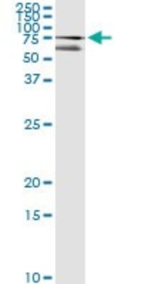
- Immunoprecipitation: CDP/CUTL1 Antibody (2A10) [H00001523-M01] - Analysis of CUX1 transfected lysate using anti-CUX1 monoclonal antibody and Protein A Magnetic Bead, and immunoblotted with CUX1 MaxPab rabbit polyclonal antibody.
Supportive validation
- Submitted by
- Novus Biologicals (provider)
- Main image
- Experimental details
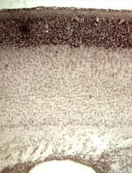
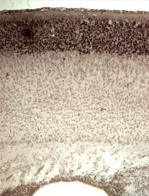
- Immunohistochemistry-Paraffin: CDP/CUTL1 Antibody (2A10) [H00001523-M01] - mouse cortex - postnatal brain was stained with anti-CUTL1 antibody. Image from verified customer review.
- Submitted by
- Novus Biologicals (provider)
- Main image
- Experimental details
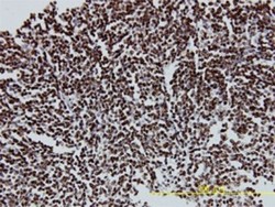
- Immunohistochemistry-Paraffin: CDP/CUTL1 Antibody (2A10) [H00001523-M01] - Analysis of monoclonal antibody to CUTL1 on formalin-fixed paraffin-embedded human malignant lymphoma, diffuse large B tissue. Antibody concentration 5 ug/ml.
