Explore
 Validate
 Learn
LearnNBP2-24397
antibody from Novus Biologicals
Targeting: FOXP3
AIID, DIETER, IPEX, JM2, PIDX, SCURFIN, XPID
 Western blot
Antibody data
- Antibody Data
- Antigen structure
- References [1]
- Comments [0]
- Validations
- Western blot [1]
- Immunohistochemistry [1]
Submit
Validation data
Reference
Comment
Report error
- Product number
- NBP2-24397 - Provider product page

- Provider
- Novus Biologicals
- Product name
- Rabbit Polyclonal FoxP3 Antibody
- Antibody type
- Polyclonal
- Description
- Protein G purified.
- Reactivity
- Human, Mouse, Rat, Bovine, Canine
- Host
- Rabbit
- Isotype
- IgG
- Vial size
- 0.1 mg
- Concentration
- 1.0 mg/ml
- Storage
- Store at 4C short term. Aliquot and store at -20C long term. Avoid freeze-thaw cycles.
Submitted references Regulatory allospecific T cell clones abrogate chronic allograft rejection.
Waaga-Gasser AM, Grimm MR, Lutz J, Lange V, Lenhard SM, Aviles B, Kist-van Holthe JE, Lebedeva T, Samsonov D, Meyer D, Hancock WW, Heemann U, Gasser M, Chandraker A
Journal of the American Society of Nephrology : JASN 2009 Apr;20(4):820-30
Journal of the American Society of Nephrology : JASN 2009 Apr;20(4):820-30
No comments: Submit comment
Supportive validation
- Submitted by
- Novus Biologicals (provider)
- Main image

- Experimental details
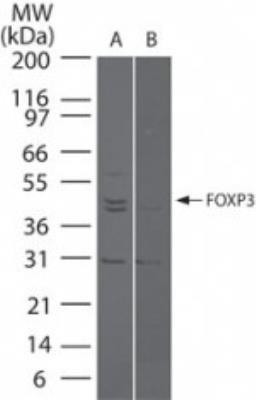
- Western Blot: FoxP3 Antibody [NBP2-24397] - Analysis of FOXP3 in (A) FOXP3 transfected and (B) mock transfected cell lysate using NBP2-24397 at 5 ug/ml.
Supportive validation
- Submitted by
- Novus Biologicals (provider)
- Main image
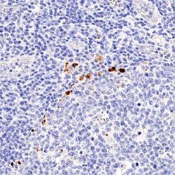
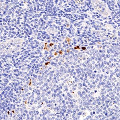
- Experimental details
- Immunohistochemistry-Paraffin: FoxP3 Antibody [NBP2-24397] - IHC analysis of formalin fixed paraffin-embedded (FFPE) human tonsil using FOXP3 antibody at 1:50 on a Bond Rx autostainer (Leica Biosystems). The assay involved 20 minutes of heat induced antigen retrieval (HIER) using 10mM sodium citrate buffer (pH 6.0) and endogenous peroxidase quenching with peroxide block. The sections were incubated with primary antibody for 30 minutes and Bond Polymer Refine Detection (Leica Biosystems) with DAB was used for signal development followed by counterstaining with hematoxylin. Whole slide scanning and capturing of representative images was performed using Aperio AT2 (Leica Biosystems). Nuclear staining was observed. Staining was performed by Histowiz.