Explore
 Validate
 Learn
LearnMA5-18160
antibody from Invitrogen Antibodies
Targeting: FOXP3
AIID, DIETER, IPEX, JM2, PIDX, SCURFIN, XPID
 Western blot
 Flow cytometry
Antibody data
- Antibody Data
- Antigen structure
- References [2]
- Comments [0]
- Validations
- Flow cytometry [4]
- Other assay [1]
Submit
Validation data
Reference
Comment
Report error
- Product number
- MA5-18160 - Provider product page

- Provider
- Invitrogen Antibodies
- Product name
- FOXP3 Monoclonal Antibody (3G3), Alexa Fluor™ 647
- Antibody type
- Monoclonal
- Antigen
- Recombinant full-length protein
- Description
- This antibody recognizes N-terminal region of FoxP3, a 47-55 kDa transcription factor (intracellular antigen), which is the master regulator in the development and function of regulatory T cells.
- Reactivity
- Human, Mouse
- Host
- Mouse
- Conjugate
- Red dye
- Isotype
- IgG
- Antibody clone number
- 3G3
- Vial size
- 100 µg
- Concentration
- 0.5 mg/mL
- Storage
- 4° C, store in dark, DO NOT FREEZE!
Submitted references A molybdenum oxide-based degradable nanosheet for combined chemo-photothermal therapy to improve tumor immunosuppression and suppress distant tumors and lung metastases.
Type I Interferons Suppress Anti-parasitic Immunity and Can Be Targeted to Improve Treatment of Visceral Leishmaniasis.
Qiu N, Yang X, Zhang Y, Zhang J, Ji J, Zhang Y, Kong X, Xi Y, Liu D, Ye L, Zhai G
Journal of nanobiotechnology 2021 Dec 19;19(1):428
Journal of nanobiotechnology 2021 Dec 19;19(1):428
Type I Interferons Suppress Anti-parasitic Immunity and Can Be Targeted to Improve Treatment of Visceral Leishmaniasis.
Kumar R, Bunn PT, Singh SS, Ng SS, Montes de Oca M, De Labastida Rivera F, Chauhan SB, Singh N, Faleiro RJ, Edwards CL, Frame TCM, Sheel M, Austin RJ, Lane SW, Bald T, Smyth MJ, Hill GR, Best SE, Haque A, Corvino D, Waddell N, Koufariotis L, Mukhopadhay P, Rai M, Chakravarty J, Singh OP, Sacks D, Nylen S, Uzonna J, Sundar S, Engwerda CR
Cell reports 2020 Feb 25;30(8):2512-2525.e9
Cell reports 2020 Feb 25;30(8):2512-2525.e9
No comments: Submit comment
Supportive validation
- Submitted by
- Invitrogen Antibodies (provider)
- Main image

- Experimental details
- Flow cytometry analysis of FOXP3 in human peripheral blood cells (gated on CD4+ cells) using FoxP3 monoclonal antibody (Product # MA5-18160).
- Conjugate
- Red dye
- Submitted by
- Invitrogen Antibodies (provider)
- Main image
- Experimental details
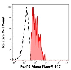
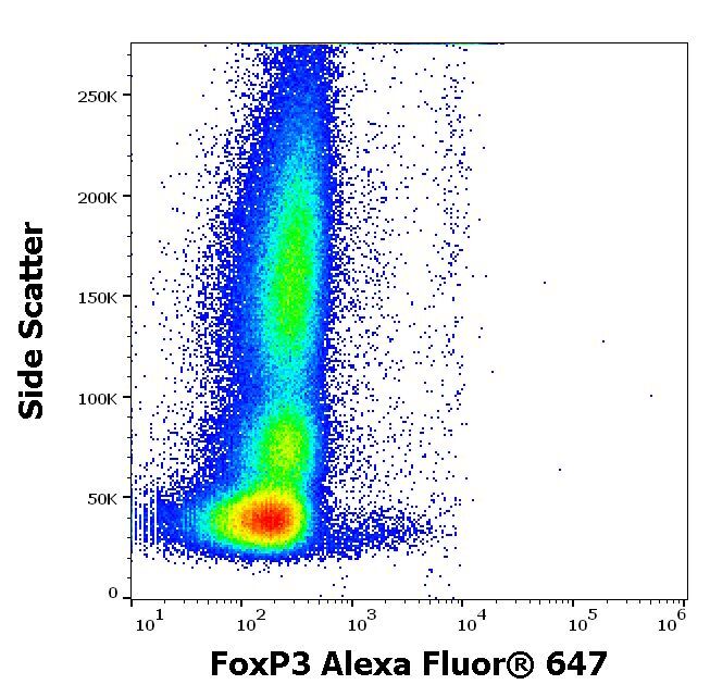
- Separation of FoxP3 positive CD25 positive CD4 positive lymphocytes (red-filled) from FoxP3 negative CD25 negative CD4 positive lymphocytes (black-dashed) in flow cytometry analysis (intracellular staining) of human peripheral whole blood stained using anti-human FoxP3 (3G3) Alexa Fluor® 647 Monoclonal antibody (Product # MA5-18160), concentration in sample 3 µg/mL.
- Conjugate
- Red dye

- Submitted by
- Invitrogen Antibodies (provider)
- Main image
- Experimental details
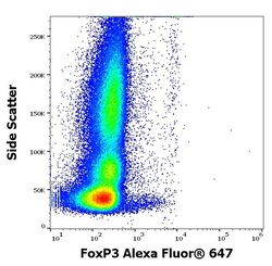
- Flow cytometry intracellular staining pattern of human peripheral whole blood stained using anti-human FoxP3 (3G3) Alexa Fluor® 647Monoclonal antibody (Product # MA5-18160), concentration in sample 3 µg/mL.
- Conjugate
- Red dye
- Submitted by
- Invitrogen Antibodies (provider)
- Main image

- Experimental details
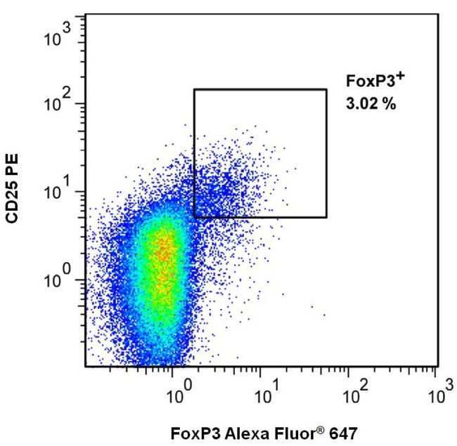
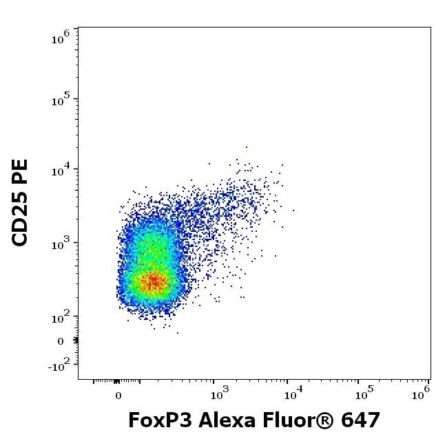
- Flow cytometry multicolor surface staining of human CD4 positive lymphocytes using anti-human CD25 (MEM-181) PE antibody (20 µL reagent / 100 µL of peripheral whole blood) and intracellular staining of human CD4 positive lymphocytes using anti-human FoxP3 (3G3) Alexa Fluor® 647 Monoclonal antibody (Product # MA5-18160), concentration in sample 3 µg/mL.
- Conjugate
- Red dye
Supportive validation
- Submitted by
- Invitrogen Antibodies (provider)
- Main image
- Experimental details
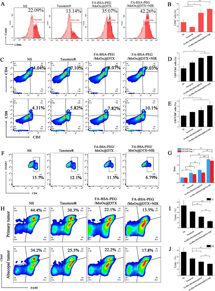
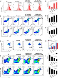
- Fig. 11 The mechanism of distant antitumor effect analysis. The flow cytometry images of DCs (CD80 + CD86 + ) mature ( A ) and quantitative analysis ( B ); flow cytometry ( C ) and quantitative analysis ( D , E ) of activated CD3 + CD4 + CD8 + T cells after each treatment; the population of CD4 + Foxp3 + Tregs in the spleen according to flow cytometry ( F ) and the ratio of CD3 + CD8 + cytotoxic T cells versus CD3 + CD4 + Foxp3 + Tregs ( G ); flow cytometry analysis of CD45 + , F4/80 + ( H ) and quantitative analysis ( I , J ) in the distant tumor
- Conjugate
- Red dye