Explore
 Validate
 Learn
Learn Immunocytochemistry
Antibody data
- Antibody Data
- Antigen structure
- References [2]
- Comments [0]
- Validations
- Immunocytochemistry [1]
- Immunohistochemistry [2]
- Flow cytometry [2]
- Other assay [1]
Submit
Validation data
Reference
Comment
Report error
- Product number
- MAB8214 - Provider product page

- Provider
- R&D Systems
- Product name
- Human/Mouse FoxP3 Antibody
- Antibody type
- Monoclonal
- Description
- Protein A or G purified from cell culture supernatant. Detects human FoxP3 in direct ELISAs. Detects human and mouse FoxP3 in flow cytometry.
- Reactivity
- Human, Mouse
- Host
- Rabbit
- Conjugate
- Unconjugated
- Antigen sequence
Q9BZS1- Isotype
- IgG
- Antibody clone number
- 1054C
- Vial size
- 100 ug
- Storage
- Use a manual defrost freezer and avoid repeated freeze-thaw cycles. 12 months from date of receipt, -20 to -70 °C as supplied. 1 month, 2 to 8 °C under sterile conditions after reconstitution. 6 months, -20 to -70 °C under sterile conditions after reconstitution.
Submitted references Improved Multiplex Immunohistochemistry for Immune Microenvironment Evaluation of Mouse Formalin-Fixed, Paraffin-Embedded Tissues.
CD25 and TGF-β blockade based on predictive integrated immune ratio inhibits tumor growth in pancreatic cancer.
Sorrelle N, Ganguly D, Dominguez ATA, Zhang Y, Huang H, Dahal LN, Burton N, Ziemys A, Brekken RA
Journal of immunology (Baltimore, Md. : 1950) 2019 Jan 1;202(1):292-299
Journal of immunology (Baltimore, Md. : 1950) 2019 Jan 1;202(1):292-299
CD25 and TGF-β blockade based on predictive integrated immune ratio inhibits tumor growth in pancreatic cancer.
Pu N, Zhao G, Yin H, Li JA, Nuerxiati A, Wang D, Xu X, Kuang T, Jin D, Lou W, Wu W
Journal of translational medicine 2018 Oct 25;16(1):294
Journal of translational medicine 2018 Oct 25;16(1):294
No comments: Submit comment
Supportive validation
- Submitted by
- R&D Systems (provider)
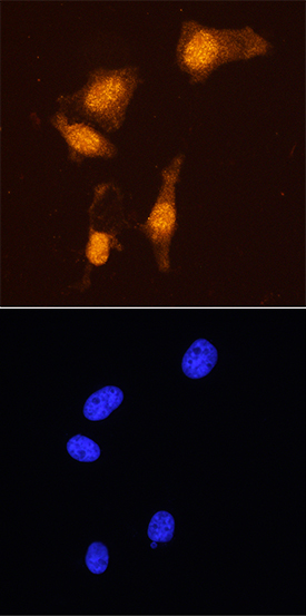
- Main image
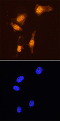
- Experimental details
- FoxP3 in HeLa Human Cell Line. FoxP3 was detected in immersion fixed HeLa human cervical epithelial carcinoma cell line using Rabbit Anti-Human/Mouse FoxP3 Monoclonal Antibody (Catalog # MAB8214) at 8 μg/mL for 3 hours at room temperature. Cells were stained using the NorthernLights™ 557-conjugated Anti-Rabbit IgG Secondary Antibody (red, upper panel; Catalog # NL004) and counterstained with DAPI (blue, lower panel). Specific staining was localized to nuclei. View our protocol for Fluorescent ICC Staining of Cells on Coverslips.
Supportive validation
- Submitted by
- R&D Systems (provider)
- Main image
- Experimental details
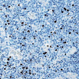
- FoxP3 in Human Tonsil. FoxP3 was detected in immersion fixed paraffin-embedded sections of human tonsil using Rabbit Anti-Human/Mouse FoxP3 Monoclonal Antibody (Catalog # MAB8214) at 15 μg/mL overnight at 4 °C. Tissue was stained using the Anti-Rabbit HRP-DAB Cell & Tissue Staining Kit (brown; Catalog # CTS005) and counterstained with hematoxylin (blue). Specific staining was localized to nuclei. View our protocol for Chromogenic IHC Staining of Paraffin-embedded Tissue Sections.
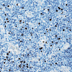
- Submitted by
- R&D Systems (provider)
- Main image
- Experimental details
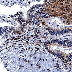
- FoxP3 in Human Ovarian Cancer Tissue. FoxP3 was detected in immersion fixed paraffin-embedded sections of human ovarian cancer tissue using Rabbit Anti-Human/Mouse FoxP3 Monoclonal Antibody (Catalog # MAB8214) at 5 µg/mL for 1 hour at room temperature followed by incubation with the Anti-Rabbit IgG VisUCyte™ HRP Polymer Antibody (Catalog # VC003). Tissue was stained using DAB (brown) and counterstained with hematoxylin (blue). Specific staining was localized to nuclei. View our protocol for IHC Staining with VisUCyte HRP Polymer Detection Reagents.
Supportive validation
- Submitted by
- R&D Systems (provider)
- Main image

- Experimental details
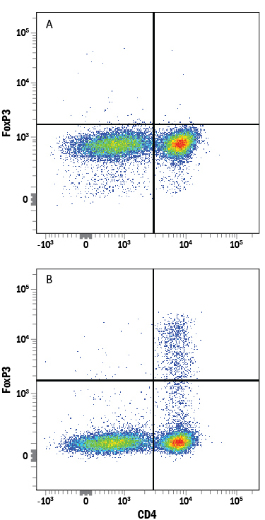
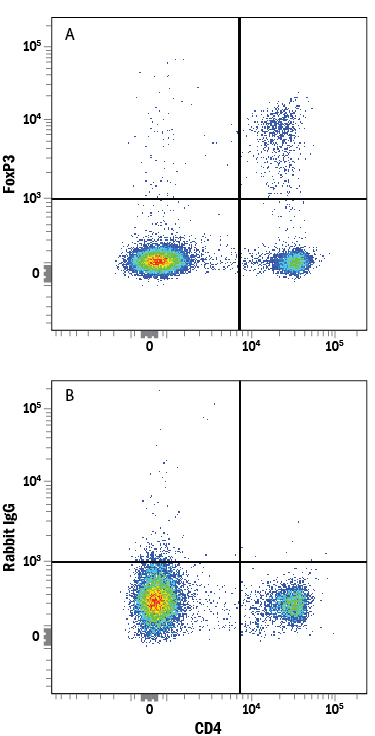
- Detection of FoxP3 in Human PBMCs stimulated to induce Tregs by Flow Cytometry. Human peripheral blood mononuclear cells (PBMCs) either (A) untreated or (B) stimulated to induce Regulatory T Cells (Tregs) with Recombinant Human TGF-beta 1 (Catalog # 240-B) and Recombinant Human IL-2 (Catalog # 202-IL) for 2 days were stained with Rabbit Anti-Human/Mouse FoxP3 Monoclonal Antibody (Catalog # MAB8214) followed by Phycoerythrin-conjugated Anti-Rabbit IgG Secondary Antibody (Catalog # F0110) and Mouse Anti-Human CD4 APC-conjugated Monoclonal Antibody (Catalog # FAB3791A). Quadrant markers were set based on control antibody staining (Catalog # AB-105-C). To facilitate intracellular staining, cells were fixed and permeabilized with FlowX FoxP3 Fixation & Permeabilization Buffer Kit (Catalog # FC012).
- Submitted by
- R&D Systems (provider)
- Main image
- Experimental details
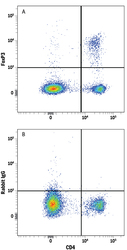
- Detection of FoxP3 in Mouse Splenocytes by Flow Cytometry. Mouse splenocytes were stained with Rat Anti-Mouse CD4 APC-conjugated Monoclonal Antibody (Catalog # FAB554A) and either (A) Rabbit Anti-Human/Mouse FoxP3 Monoclonal Antibody (Catalog # MAB8214) or (B) Normal Rabbit IgG Control (Catalog # AB-105-C) followed by Phycoerythrin-conjugated Anti-Rabbit IgG Secondary Antibody (Catalog # F0110). To facilitate intracellular staining, cells were fixed and permeabilized with FlowX FoxP3 Fixation & Permeabilization Buffer Kit (Catalog # FC012).
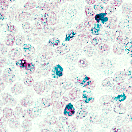
Supportive validation
- Submitted by
- R&D Systems (provider)
- Main image
- Experimental details
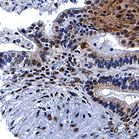
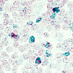
- FoxP3 in Human Tonsil Using Dual RNAscope® ISH and IHC. FoxP3 mRNA (red) and protein (green) was detected in formalin-fixed paraffin-embedded tissue sections of human tonsil probed with ACD RNAScope® Probe (Catalog # 418471) followed by immunohistochemistry using R&D Systems Rabbit Anti-Human/Mouse FoxP3 Monoclonal Antibody (Catalog# MAB8214) at 5ug/mL for 1 hour at room temperature followed by incubation with the Anti-Rabbit IgG VisUCyte HRP Polymer Antibody (R&D Systems, Catalog # VC003). Tissue was stained using ACD RNAscope® 2.5 HD Duplex Detection Reagents (Catalog # 322500).